Journal of Molecular and Cellular Cardiology ( IF 4.9 ) Pub Date : 2020-08-12 , DOI: 10.1016/j.yjmcc.2020.08.005 Valérie Long 1 , Céline Fiset 1
|
Background
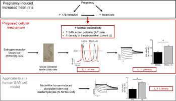
The heart rate progressively increases throughout pregnancy, reaching a maximum in the third trimester. This elevated heart rate is also present in pregnant mice and is associated with accelerated automaticity, higher density of the pacemaker current If and changes in Ca2+ homeostasis in sinoatrial node (SAN) cells. Strong evidence has also been provided showing that 17β-estradiol (E2) and estrogen receptor α (ERα) regulate heart rate. Accordingly, we sought to determine whether E2 levels found in late pregnancy cause the increased cardiac automaticity associated with pregnancy.
Methods and results
Voltage- and current-clamp experiments were carried out on SAN cells isolated from female mice lacking estrogen receptor alpha (ERKOα) or beta (ERKOβ) receiving chronic E2 treatment mimicking late pregnancy concentrations. E2 treatment significantly increased the action potential rate (284 ± 24 bpm, +E2 354 ± 23 bpm, p = 0.040) and the density of If (+52%) in SAN cells from ERKOβ mice. However, If density remains unchanged in SAN cells from E2-treated ERKOα mice. Additionally, E2 also increased If density (+67%) in nodal-like human-induced pluripotent stem cell-derived cardiomyocytes (N-hiPSC-CM), recapitulating in a human SAN cell model the effect produced in mice. However, the L-type calcium current (ICaL) and Ca2+ transients, examined using N-hiPSC-CM and SAN cells respectively, were not affected by E2, indicating that other mechanisms contribute to changes observed in these parameters during pregnancy.
Conclusion
The accelerated SAN automaticity observed in E2-treated ERKOβ mice is explained by an increased If density mediated by ERα, demonstrating that E2 plays a major role in regulating SAN function during pregnancy.
中文翻译:
雌激素对妊娠引起的心脏自动性增加的贡献。
背景
在整个怀孕期间,心率逐渐增加,在孕晚期达到最高。这种心率升高也存在于怀孕的小鼠中,并且与加速的自动化,起搏器电流I f的更高密度以及窦房结(SAN)细胞中Ca 2+稳态的变化有关。还提供了强有力的证据,表明17β-雌二醇(E 2)和雌激素受体α(ERα)调节心率。因此,我们试图确定在妊娠晚期发现的E 2水平是否会导致与妊娠相关的心脏自动性增加。
方法与结果
在从缺乏雌激素受体α(ERKOα)或β(ERKOβ)的雌性小鼠中分离的SAN细胞上进行了电压和电流钳实验,这些小鼠接受了模拟晚期妊娠浓度的慢性E 2治疗。E 2处理显着提高了ERKOβ小鼠SAN细胞中的动作电位率(284±24 bpm,+ E 2 354±23 bpm,p = 0.040)和I f(+ 52%)密度。然而,在E 2处理的ERKOα小鼠的SAN细胞中,I f密度保持不变。此外,E 2也增加了I f节点样人类诱导的多能干细胞衍生的心肌细胞(N-hiPSC-CM)中的密度(+ 67%),在人类SAN细胞模型中概括了小鼠产生的效应。然而,分别使用N-hiPSC-CM和SAN细胞检查的L型钙电流(I CaL)和Ca 2+瞬变不受E 2的影响,表明在怀孕期间这些参数中观察到的其他机制也有变化。
结论
在E 2处理的ERKOβ小鼠中观察到的SAN自动化加速是由ERα介导的I f密度增加所解释的,这表明E 2在孕期调节SAN功能中起主要作用。


 京公网安备 11010802027423号