Journal of Structural Biology ( IF 3.0 ) Pub Date : 2020-08-09 , DOI: 10.1016/j.jsb.2020.107598 Natalie Reznikov 1 , Dan J Buss 2 , Benjamin Provencher 1 , Marc D McKee 3 , Nicolas Piché 1
|
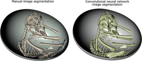
Biomineralization research examines structure-function relations in all types of exo- and endo-skeletons and other hard tissues of living organisms, and it relies heavily on 3D imaging. Segmentation of 3D renderings of biomineralized structures has long been a bottleneck because of human limitations such as our available time, attention span, eye-hand coordination, cognitive biases, and attainable precision, amongst other limitations. Since recently, some of these routine limitations appear to be surmountable thanks to the development of deep-learning algorithms for biological imagery in general, and for 3D image segmentation in particular. Many components of deep learning often appear too abstract for a life scientist. Despite this, the basic principles underlying deep learning have many easy-to-grasp commonalities with human learning and universal logic. This primer presents these basic principles in what we feel is an intuitive manner, without relying on prerequisite knowledge of informatics and computer science, and with the aim of improving the reader’s general literacy in artificial intelligence and deep learning. Here, biomineralization case studies are presented to illustrate the application of deep learning for solving segmentation and analysis problems of 3D images ridden by various artifacts, and/or which are plainly difficult to interpret. The presented portfolio of case studies includes three examples of imaging using micro-computed tomography (µCT), and three examples using focused-ion beam scanning electron microscopy (FIB-SEM), all on mineralized tissues. We believe this primer will expand the circle of users of deep learning amongst biomineralization researchers and other life scientists involved with 3D imaging, and will encourage incorporation of this powerful tool into their professional skillsets and to explore it further.
中文翻译:
生物矿化研究中 3D 成像和图像分析的深度学习。
生物矿化研究检查了所有类型的外型和内型的结构 - 功能关系- 生物体的骨骼和其他硬组织,它严重依赖 3D 成像。由于人类的限制,例如我们的可用时间、注意力跨度、眼手协调、认知偏差和可达到的精度,以及其他限制,生物矿化结构的 3D 渲染的分割长期以来一直是一个瓶颈。最近,由于开发了用于一般生物图像,特别是 3D 图像分割的深度学习算法,这些常规限制中的一些似乎可以克服。对于生命科学家来说,深度学习的许多组成部分通常显得过于抽象。尽管如此,深度学习的基本原理与人类学习和通用逻辑有许多易于掌握的共性。本入门书以我们认为直观的方式介绍了这些基本原则,不依赖信息学和计算机科学的必备知识,旨在提高读者在人工智能和深度学习方面的综合素养。在这里,生物矿化案例研究展示了深度学习在解决由各种伪影和/或明显难以解释的 3D 图像的分割和分析问题中的应用。所呈现的案例研究组合包括三个使用显微计算机断层扫描 (µCT) 成像的示例,以及三个使用聚焦离子束扫描电子显微镜 (FIB-SEM) 的示例,所有这些都在矿化组织上进行。我们相信这本入门书将扩大生物矿化研究人员和其他参与 3D 成像的生命科学家的深度学习用户圈,


 京公网安备 11010802027423号