当前位置:
X-MOL 学术
›
J. Neurosci. Res.
›
论文详情
Our official English website, www.x-mol.net, welcomes your
feedback! (Note: you will need to create a separate account there.)
Melatonin alters neuronal architecture and increases cysteine-rich protein 1 signaling in the male mouse hippocampus.
Journal of Neuroscience Research ( IF 2.9 ) Pub Date : 2020-08-04 , DOI: 10.1002/jnr.24708 Mary Jasmin Ang 1 , Sohi Kang 1 , Changjong Moon 1
Journal of Neuroscience Research ( IF 2.9 ) Pub Date : 2020-08-04 , DOI: 10.1002/jnr.24708 Mary Jasmin Ang 1 , Sohi Kang 1 , Changjong Moon 1
Affiliation
|
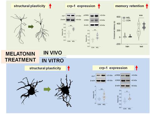
Neuronal plasticity describes changes in structure, function, and connections of neurons. The hippocampus, in particular, has been shown to exhibit considerable plasticity regarding both physiological and morphological functions. Melatonin, a hormone released by the pineal gland, promotes cell survival and dendrite maturation of neurons in the newborn brain and protects against neurological disorders. In this study, we investigated the effect of exogenous melatonin on neuronal architecture and its possible mechanism in the hippocampus of adult male C57BL/6 mice. Melatonin treatment significantly increased the total length and complexity of dendrites in the apical and basal cornu ammonis (CA) 1 and in the dentate gyrus in mouse hippocampi. Spine density in CA1 apical dendrites was increased, but no significant differences in other subregions were observed. In primary cultured hippocampal neurons, the length and arborization of neurites were significantly augmented by melatonin treatment. Additionally, western blot and immunohistochemical analyses in both in vivo and in vitro systems revealed significant increases in the level of cysteine‐rich protein 1 (crp‐1) protein, which is known to be involved in dendritic branching in mouse hippocampal neurons after melatonin treatment. Our results suggest that exogenous melatonin leads to significant alterations of neuronal micromorphometry in the adult hippocampus, possibly via crp‐1 signaling.
中文翻译:

褪黑激素改变神经元结构并增加雄性小鼠海马体中富含半胱氨酸的蛋白 1 信号。
神经元可塑性描述了神经元结构、功能和连接的变化。特别是海马体已被证明在生理和形态功能方面表现出相当大的可塑性。褪黑激素是松果体释放的一种激素,可促进新生大脑中神经元的细胞存活和树突成熟,并防止神经系统疾病。在这项研究中,我们研究了外源性褪黑激素对成年雄性 C57BL/6 小鼠海马神经元结构的影响及其可能的机制。褪黑激素治疗显着增加了根尖和基底角树突的总长度和复杂性(CA) 1 和小鼠海马的齿状回。CA1 顶端树突的脊椎密度增加,但未观察到其他亚区域的显着差异。在原代培养的海马神经元中,褪黑激素处理显着增加了神经突的长度和分支。此外,体内和体外系统中的蛋白质印迹和免疫组织化学分析显示富含半胱氨酸的蛋白 1 (crp-1) 蛋白水平显着增加,已知该蛋白在褪黑激素治疗后参与小鼠海马神经元的树突分支. 我们的结果表明,外源性褪黑激素可能通过 crp-1 信号传导导致成年海马神经元微形态测量的显着改变。
更新日期:2020-10-04
中文翻译:
褪黑激素改变神经元结构并增加雄性小鼠海马体中富含半胱氨酸的蛋白 1 信号。
神经元可塑性描述了神经元结构、功能和连接的变化。特别是海马体已被证明在生理和形态功能方面表现出相当大的可塑性。褪黑激素是松果体释放的一种激素,可促进新生大脑中神经元的细胞存活和树突成熟,并防止神经系统疾病。在这项研究中,我们研究了外源性褪黑激素对成年雄性 C57BL/6 小鼠海马神经元结构的影响及其可能的机制。褪黑激素治疗显着增加了根尖和基底角树突的总长度和复杂性(CA) 1 和小鼠海马的齿状回。CA1 顶端树突的脊椎密度增加,但未观察到其他亚区域的显着差异。在原代培养的海马神经元中,褪黑激素处理显着增加了神经突的长度和分支。此外,体内和体外系统中的蛋白质印迹和免疫组织化学分析显示富含半胱氨酸的蛋白 1 (crp-1) 蛋白水平显着增加,已知该蛋白在褪黑激素治疗后参与小鼠海马神经元的树突分支. 我们的结果表明,外源性褪黑激素可能通过 crp-1 信号传导导致成年海马神经元微形态测量的显着改变。


 京公网安备 11010802027423号