当前位置:
X-MOL 学术
›
Mol. Reprod. Dev.
›
论文详情
Our official English website, www.x-mol.net, welcomes your
feedback! (Note: you will need to create a separate account there.)
Characterization of rodent Sertoli cell primary cultures.
Molecular Reproduction and Development ( IF 2.7 ) Pub Date : 2020-08-02 , DOI: 10.1002/mrd.23402 Helena D Zomer 1 , Prabhakara P Reddi 1
Molecular Reproduction and Development ( IF 2.7 ) Pub Date : 2020-08-02 , DOI: 10.1002/mrd.23402 Helena D Zomer 1 , Prabhakara P Reddi 1
Affiliation
|
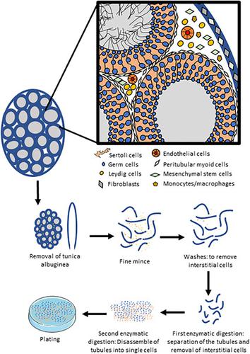
Sertoli cells play a vital role in spermatogenesis by offering physical and nutritional support to the differentiating male germ cells. They form the blood–testis barrier and secrete growth factors essential for germ cell differentiation. Sertoli cell primary cultures are critical for understanding the regulation of spermatogenesis; however, obtaining pure cultures has been a challenge. Rodent Sertoli cell isolation protocols do not rule out contamination by the interstitial or connective tissue cells. Sertoli cell‐specific markers could be helpful, but there is no consensus. Vimentin, the most commonly used marker, is not specific for Sertoli cells since its expression has been reported in peritubular myoid cells, mesenchymal stem cells, fibroblasts, macrophages, and endothelial cells, which contaminate Sertoli cell preparations. Markers based on transcription and growth factors also have limitations. Thus, the impediment to obtaining pure Sertoli cell cultures pertains to both the method of isolation and marker usage. The aim of this review is to discuss improvements to current methods of rodent Sertoli cell primary cultures, assess the properties of prepubertal versus mature Sertoli cell cultures, and propose steps to improve cellular characterization. Potential benefits of using contemporary approaches, including lineage tracing, specific cell ablation, and RNA‐seq for obtaining Sertoli‐specific transcript markers are discussed. Evaluating the specificity and applicability of these markers at the protein level to characterize Sertoli cells in culture would be critical. This review is expected to positively impact future work using primary cultures of rodent Sertoli cells.
中文翻译:

啮齿动物支持细胞原代培养物的表征。
支持细胞为分化的雄性生殖细胞提供物理和营养支持,在精子发生中发挥着至关重要的作用。它们形成血睾屏障并分泌生殖细胞分化所必需的生长因子。支持细胞原代培养对于理解精子发生的调节至关重要;然而,获得纯培养物一直是一个挑战。啮齿动物支持细胞分离方案不排除间质或结缔组织细胞的污染。支持细胞特异性标记物可能有所帮助,但尚未达成共识。波形蛋白是最常用的标记物,它对支持细胞并不具有特异性,因为据报道其在管周肌样细胞、间充质干细胞、成纤维细胞、巨噬细胞和内皮细胞中表达,这些细胞会污染支持细胞制剂。基于转录和生长因子的标记也有局限性。因此,获得纯支持细胞培养物的障碍与分离方法和标记物的使用有关。本综述的目的是讨论对当前啮齿类支持细胞原代培养方法的改进,评估青春期前支持细胞培养与成熟支持细胞培养的特性,并提出改善细胞特征的步骤。讨论了使用现代方法(包括谱系追踪、特定细胞消融和 RNA 测序)获得支持细胞特异性转录标记物的潜在好处。在蛋白质水平上评估这些标记物的特异性和适用性以表征培养物中支持细胞的特征至关重要。该综述预计将对未来使用啮齿动物支持细胞原代培养物的工作产生积极影响。
更新日期:2020-08-28
中文翻译:
啮齿动物支持细胞原代培养物的表征。
支持细胞为分化的雄性生殖细胞提供物理和营养支持,在精子发生中发挥着至关重要的作用。它们形成血睾屏障并分泌生殖细胞分化所必需的生长因子。支持细胞原代培养对于理解精子发生的调节至关重要;然而,获得纯培养物一直是一个挑战。啮齿动物支持细胞分离方案不排除间质或结缔组织细胞的污染。支持细胞特异性标记物可能有所帮助,但尚未达成共识。波形蛋白是最常用的标记物,它对支持细胞并不具有特异性,因为据报道其在管周肌样细胞、间充质干细胞、成纤维细胞、巨噬细胞和内皮细胞中表达,这些细胞会污染支持细胞制剂。基于转录和生长因子的标记也有局限性。因此,获得纯支持细胞培养物的障碍与分离方法和标记物的使用有关。本综述的目的是讨论对当前啮齿类支持细胞原代培养方法的改进,评估青春期前支持细胞培养与成熟支持细胞培养的特性,并提出改善细胞特征的步骤。讨论了使用现代方法(包括谱系追踪、特定细胞消融和 RNA 测序)获得支持细胞特异性转录标记物的潜在好处。在蛋白质水平上评估这些标记物的特异性和适用性以表征培养物中支持细胞的特征至关重要。该综述预计将对未来使用啮齿动物支持细胞原代培养物的工作产生积极影响。


 京公网安备 11010802027423号