当前位置:
X-MOL 学术
›
J. Neurosci. Res.
›
论文详情
Our official English website, www.x-mol.net, welcomes your
feedback! (Note: you will need to create a separate account there.)
Toward quantitative neuroimaging biomarkers for Friedreich's ataxia at 7 Tesla: Susceptibility mapping, diffusion imaging, R2 and R1 relaxometry.
Journal of Neuroscience Research ( IF 2.9 ) Pub Date : 2020-07-30 , DOI: 10.1002/jnr.24701 Sina Straub 1 , Stephanie Mangesius 2, 3 , Julian Emmerich 1, 4 , Elisabetta Indelicato 5 , Wolfgang Nachbauer 5 , Katja S Degenhardt 1, 4 , Mark E Ladd 1, 4, 6 , Sylvia Boesch 5 , Elke R Gizewski 2, 3
Journal of Neuroscience Research ( IF 2.9 ) Pub Date : 2020-07-30 , DOI: 10.1002/jnr.24701 Sina Straub 1 , Stephanie Mangesius 2, 3 , Julian Emmerich 1, 4 , Elisabetta Indelicato 5 , Wolfgang Nachbauer 5 , Katja S Degenhardt 1, 4 , Mark E Ladd 1, 4, 6 , Sylvia Boesch 5 , Elke R Gizewski 2, 3
Affiliation
|
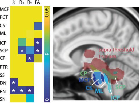
Friedreich's ataxia (FRDA) is a rare genetic disorder leading to degenerative processes. So far, no effective treatment has been found. Therefore, it is important to assist the development of medication with imaging biomarkers reflecting disease status and progress. Ten FRDA patients (mean age 37 ± 14 years; four female) and 10 age‐ and sex‐matched controls were included. Acquisition of magnetic resonance imaging (MRI) data for quantitative susceptibility mapping, R1, R2 relaxometry and diffusion imaging was performed at 7 Tesla. Results of volume of interest (VOI)‐based analyses of the quantitative data were compared with a voxel‐based morphometry (VBM) evaluation. Differences between patients and controls were assessed using the analysis of covariance (ANCOVA; p < 0.01) with age and sex as covariates, effect size of group differences, and correlations with disease characteristics with Spearman correlation coefficient. For the VBM analysis, a statistical threshold of 0.001 for uncorrected and 0.05 for corrected p‐values was used. Statistically significant differences between FRDA patients and controls were found in five out of twelve investigated structures, and statistically significant correlations with disease characteristics were revealed. Moreover, VBM revealed significant white matter atrophy within regions of the brainstem, and the cerebellum. These regions overlapped partially with brain regions for which significant differences between healthy controls and patients were found in the VOI‐based quantitative MRI evaluation. It was shown that two independent analyses provided overlapping results. Moreover, positive results on correlations with disease characteristics were found, indicating that these quantitative MRI parameters could provide more detailed information and assist the search for effective treatments.
中文翻译:

迈向 7 特斯拉弗里德赖希共济失调的定量神经成像生物标志物:磁化率映射、扩散成像、R2 和 R1 弛豫测量。
弗里德赖希共济失调 (FRDA) 是一种罕见的遗传性疾病,可导致退行性过程。到目前为止,还没有找到有效的治疗方法。因此,重要的是通过反映疾病状态和进展的成像生物标志物来协助药物的开发。包括 10 名 FRDA 患者(平均年龄 37 ± 14 岁;4 名女性)和 10 名年龄和性别匹配的对照。获取磁共振成像 (MRI) 数据,用于定量磁化率映射、R 1、R 2弛豫和扩散成像在 7 特斯拉进行。将基于感兴趣体积 (VOI) 的定量数据分析结果与基于体素的形态测量 (VBM) 评估进行比较。使用协方差分析 (ANCOVA;p < 0.01)评估患者和对照之间的差异,年龄和性别作为协变量,组差异的影响大小,以及与疾病特征的相关性与 Spearman 相关系数。对于 VBM 分析,未校正p的统计阈值为 0.001,校正p的统计阈值为 0.05- 值被使用。在 FRDA 患者和对照组之间的 12 个研究结构中的 5 个中发现了统计学上的显着差异,并揭示了与疾病特征的统计学显着相关性。此外,VBM 显示脑干和小脑区域内有明显的白质萎缩。这些区域与大脑区域部分重叠,在基于 VOI 的定量 MRI 评估中发现健康对照组和患者之间存在显着差异。结果表明,两个独立的分析提供了重叠的结果。此外,发现与疾病特征相关的积极结果,表明这些定量的 MRI 参数可以提供更详细的信息,并有助于寻找有效的治疗方法。
更新日期:2020-10-04
中文翻译:
迈向 7 特斯拉弗里德赖希共济失调的定量神经成像生物标志物:磁化率映射、扩散成像、R2 和 R1 弛豫测量。
弗里德赖希共济失调 (FRDA) 是一种罕见的遗传性疾病,可导致退行性过程。到目前为止,还没有找到有效的治疗方法。因此,重要的是通过反映疾病状态和进展的成像生物标志物来协助药物的开发。包括 10 名 FRDA 患者(平均年龄 37 ± 14 岁;4 名女性)和 10 名年龄和性别匹配的对照。获取磁共振成像 (MRI) 数据,用于定量磁化率映射、R 1、R 2弛豫和扩散成像在 7 特斯拉进行。将基于感兴趣体积 (VOI) 的定量数据分析结果与基于体素的形态测量 (VBM) 评估进行比较。使用协方差分析 (ANCOVA;p < 0.01)评估患者和对照之间的差异,年龄和性别作为协变量,组差异的影响大小,以及与疾病特征的相关性与 Spearman 相关系数。对于 VBM 分析,未校正p的统计阈值为 0.001,校正p的统计阈值为 0.05- 值被使用。在 FRDA 患者和对照组之间的 12 个研究结构中的 5 个中发现了统计学上的显着差异,并揭示了与疾病特征的统计学显着相关性。此外,VBM 显示脑干和小脑区域内有明显的白质萎缩。这些区域与大脑区域部分重叠,在基于 VOI 的定量 MRI 评估中发现健康对照组和患者之间存在显着差异。结果表明,两个独立的分析提供了重叠的结果。此外,发现与疾病特征相关的积极结果,表明这些定量的 MRI 参数可以提供更详细的信息,并有助于寻找有效的治疗方法。


 京公网安备 11010802027423号