Structure ( IF 4.4 ) Pub Date : 2020-07-30 , DOI: 10.1016/j.str.2020.07.006 Lan Zhu 1 , Guanhong Bu 2 , Liang Jing 1 , Dan Shi 3 , Ming-Yue Lee 1 , Tamir Gonen 4 , Wei Liu 1 , Brent L Nannenga 2
|
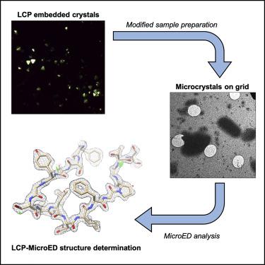
The lipidic cubic phase (LCP) technique has proved to facilitate the growth of high-quality crystals that are otherwise difficult to grow by other methods. However, the crystal size optimization process could be time and resource consuming, if it ever happens. Therefore, improved techniques for structure determination using these small crystals is an important strategy in diffraction technology development. Microcrystal electron diffraction (MicroED) is a technique that uses a cryo-transmission electron microscopy to collect electron diffraction data and determine high-resolution structures from very thin micro- and nanocrystals. In this work, we have used modified LCP and MicroED protocols to analyze crystals embedded in LCP converted by 2-methyl-2,4-pentanediol or lipase, including Proteinase K crystals grown in solution, cholesterol crystals, and human adenosine A2A receptor crystals grown in LCP. These results set the stage for the use of MicroED to analyze microcrystalline samples grown in LCP, especially for those highly challenging membrane protein targets.
中文翻译:
通过 MicroED 测定脂质立方相嵌入微晶的结构。
脂质立方相(LCP)技术已被证明可以促进高质量晶体的生长,而其他方法很难生长这些晶体。然而,晶体尺寸优化过程如果发生的话可能会耗费时间和资源。因此,利用这些小晶体改进结构测定技术是衍射技术发展的重要策略。微晶电子衍射 (MicroED) 是一种使用低温透射电子显微镜收集电子衍射数据并确定非常薄的微米和纳米晶体的高分辨率结构的技术。在这项工作中,我们使用改进的 LCP 和 MicroED 方案来分析嵌入在由 2-甲基-2,4-戊二醇或脂肪酶转化的 LCP 中的晶体,包括在溶液中生长的蛋白酶 K 晶体、胆固醇晶体和人腺苷 A 2A受体晶体在 LCP 中生长。这些结果为使用 MicroED 分析 LCP 中生长的微晶样品奠定了基础,尤其是那些极具挑战性的膜蛋白靶标。


 京公网安备 11010802027423号