Journal of Structural Biology ( IF 3.0 ) Pub Date : 2020-07-28 , DOI: 10.1016/j.jsb.2020.107592 Wenge Jiang 1 , Gabriele Griffanti 2 , Faleh Tamimi 3 , Marc D McKee 4 , Showan N Nazhat 2
|
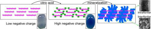
The mineralized extracellular matrix of bone is an organic–inorganic nanocomposite consisting primarily of carbonated hydroxyapatite, fibrous type I collagen, noncollagenous proteins, proteoglycans, and diverse biomolecules such as pyrophosphate and citrate. While much is now known about the mineralization-regulating role of pyrophosphate, less is known about the function of citrate. In order to assess the effect of negatively charged citrate on collagen mineralization, citrate-functionalized, bone osteoid-mimicking dense collagen gels were exposed to simulated body fluid for up to 7 days to examine the multiscale evolution of intra- and interfibrillar collagen mineralization. Here, we show by increases in methylene blue staining that the net negative charge of collagen can be substantially augmented through citrate functionalization. Structural and compositional analyses by transmission and scanning electron microscopy (including X-ray microanalysis and electron diffraction), and atomic force microscopy, all demonstrated that citrate-functionalized collagen fibrils underwent extensive intrafibrillar mineralization within 12 h in simulated body fluid. Time-resolved, high-resolution transmission electron microscopy confirmed the temporal evolution of intrafibrillar mineralization of single collagen fibrils. Longer exposure to simulated body fluid resulted in additional interfibrillar mineralization, all through an amorphous-to-crystalline transformation towards apatite (assessed by X-ray diffraction and attenuated total reflection-Fourier-transform infrared spectroscopy). Calcium deposition assays indicated a citrate concentration-dependent temporal increase in mineralization, and micro-computed tomography confirmed that >80 vol% of the collagen in the gels was mineralized by day 7. In conclusion, citrate effectively induces mesoscale intra- and interfibrillar collagen mineralization, a finding that advances our understanding of the role of citrate in mineralized tissues.
中文翻译:
致密胶原凝胶中柠檬酸盐触发的纤维内和纤维间矿化的多尺度结构演化。
骨的矿化细胞外基质是一种有机-无机纳米复合材料,主要由碳酸羟基磷灰石、纤维状 I 型胶原蛋白、非胶原蛋白、蛋白聚糖和多种生物分子(如焦磷酸盐和柠檬酸盐)组成。虽然现在人们对焦磷酸盐的矿化调节作用知之甚少,但对柠檬酸盐的功能知之甚少。为了评估带负电荷的柠檬酸盐对胶原矿化的影响,将柠檬酸盐功能化的骨类骨模拟致密胶原凝胶暴露于模拟体液中长达 7 天,以检查纤维内和纤维间胶原矿化的多尺度演变。在这里,我们通过亚甲蓝染色的增加表明胶原蛋白的净负电荷可以通过柠檬酸盐功能化显着增加。通过透射和扫描电子显微镜(包括 X 射线微量分析和电子衍射)以及原子力显微镜进行的结构和成分分析都表明,柠檬酸盐功能化的胶原纤维在模拟体液中在 12 小时内经历了广泛的纤维内矿化。时间分辨、高分辨率透射电子显微镜证实了单个胶原纤维的纤维内矿化的时间演变。长时间暴露于模拟体液导致额外的纤维间矿化,所有这些都是通过无定形到结晶向磷灰石的转变(通过 X 射线衍射和衰减全反射傅里叶变换红外光谱评估)。钙沉积测定表明柠檬酸盐浓度依赖性矿化随时间增加,


 京公网安备 11010802027423号