Photoacoustics ( IF 7.1 ) Pub Date : 2020-07-25 , DOI: 10.1016/j.pacs.2020.100201 Eno Hysi 1, 2 , Muhannad N Fadhel 1, 2 , Yanjie Wang 1, 2 , Joseph A Sebastian 1, 2 , Anoja Giles 3, 4 , Gregory J Czarnota 3, 4, 5, 6 , Agata A Exner 7, 8 , Michael C Kolios 1, 2
|
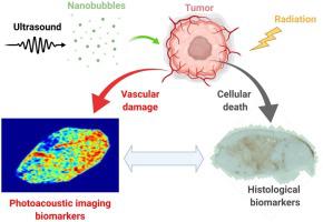
The development of novel anticancer therapies warrants the parallel development of biomarkers that can quantify their effectiveness. Photoacoustic imaging has the potential to measure changes in tumor vasculature during treatment. Establishing the accuracy of imaging biomarkers requires direct comparisons with gold histological standards. In this work, we explore whether a new class of submicron, vascular disrupting, ultrasonically stimulated nanobubbles enhance radiation therapy. In vivo experiments were conducted on mice bearing prostate cancer tumors. Combined nanobubble plus radiation treatments were compared against conventional microbubbles and radiation alone (single 8 Gy fraction). Acoustic resolution photoacoustic imaging was used to monitor the effects of the treatments 2- and 24-hs post-administration. Histological examination provided metrics of tumor vascularity and tumoral cell death, both of which were compared to photoacoustic-derived biomarkers. Photoacoustic metrics of oxygen saturation reveal a 20 % decrease in oxygenation within 24 h post-treatment. The spectral slope metric could separate the response of the nanobubble treatments from the microbubble counterparts. This study shows that histopathological assessment correlated well with photoacoustic biomarkers of treatment response.
中文翻译:
用于监测纳米气泡介导的放射治疗期间生物物理变化的光声成像生物标志物。
新型抗癌疗法的发展需要同时开发能够量化其有效性的生物标志物。光声成像有可能测量治疗期间肿瘤脉管系统的变化。确定成像生物标志物的准确性需要与黄金组织学标准进行直接比较。在这项工作中,我们探索了一类新型亚微米、血管破坏、超声波刺激纳米气泡是否可以增强放射治疗。对患有前列腺癌肿瘤的小鼠进行了体内实验。将纳米气泡加辐射组合治疗与传统微泡和单独辐射(单次 8 Gy 剂量)进行比较。使用声分辨率光声成像来监测给药后2小时和24小时的治疗效果。组织学检查提供了肿瘤血管分布和肿瘤细胞死亡的指标,将两者与光声衍生的生物标志物进行比较。氧饱和度的光声指标显示治疗后 24 小时内氧合下降 20%。光谱斜率度量可以将纳米气泡处理的响应与微气泡对应物的响应分开。这项研究表明,组织病理学评估与治疗反应的光声生物标志物密切相关。


 京公网安备 11010802027423号