当前位置:
X-MOL 学术
›
ACS Appl. Mater. Interfaces
›
论文详情
Our official English website, www.x-mol.net, welcomes your
feedback! (Note: you will need to create a separate account there.)
Enhanced biocompatibility and differentiation capacity of mesenchymal stem cells on polydimethylsiloxane by topographical patterned dopamine.
ACS Applied Materials & Interfaces ( IF 8.3 ) Pub Date : 2020-07-22 , DOI: 10.1021/acsami.0c05747 Huey-Shan Hung,Yang-Hao Yu,Shuchen Hsieh,Mei-Lang Kung,Hsiu-Yuan Huang,Ru-Huei Fu,Chun-An Yeh,Shan-Hui Hsu
ACS Applied Materials & Interfaces ( IF 8.3 ) Pub Date : 2020-07-22 , DOI: 10.1021/acsami.0c05747 Huey-Shan Hung,Yang-Hao Yu,Shuchen Hsieh,Mei-Lang Kung,Hsiu-Yuan Huang,Ru-Huei Fu,Chun-An Yeh,Shan-Hui Hsu

|
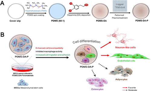
Controlling the behavior of mesenchymal stem cells (MSCs) through topographic patterns is an effective approach for stem cell studies. We, herein, reported a facile method to create a dopamine (DA) pattern on poly(dimethylsiloxane) (PDMS). The topography of micropatterned DA was produced on PDMS after plasma treatment. The grid-topographic-patterned surface of PDMS-DA (PDMS-DA-P) was measured for adhesion force and Young’s modulus by atomic force microscopy. The surface of PDMS-DA-P demonstrated less stiff and more elastic characteristics compared to either nonpatterned PDMS-DA or PDMS. The PDMS-DA-P evidently enhanced the differentiation of MSCs into various tissue cells, including nerve, vessel, bone, and fat. We further designed comprehensive experiments to investigate adhesion, proliferation, and differentiation of MSCs in response to PDMS-DA-P and showed that the DA-patterned surface had good biocompatibility and did not activate macrophages or platelets in vitro and had low foreign body reaction in vivo. Besides, it protected MSCs from apoptosis as well as excessive reactive oxygen species (ROS) generation. Particularly, the patterned surface enhanced the differentiation capacity of MSCs toward neural and endothelial cells. The stromal cell-derived factor-1α/CXantiCR4 pathway may be involved in mediating the self-recruitment and promoting the differentiation of MSCs. These findings support the potential application of PDMS-DA-P in either cell treatment or tissue repair.
中文翻译:

地形图式多巴胺增强了间充质干细胞在聚二甲基硅氧烷上的生物相容性和分化能力。
通过地形图模式控制间充质干细胞(MSC)的行为是干细胞研究的有效方法。我们在本文中报道了一种在聚二甲基硅氧烷(PDMS)上创建多巴胺(DA)模式的简便方法。等离子体处理后,在PDMS上产生了微图案DA的形貌。通过原子力显微镜测量PDMS-DA(PDMS-DA-P)的网格地形图表面的粘附力和杨氏模量。与未图案化的PDMS-DA或PDMS相比,PDMS-DA-P的表面显示出较少的硬度和弹性。PDMS-DA-P明显增强了MSCs向各种组织细胞的分化,包括神经,血管,骨骼和脂肪。我们进一步设计了综合实验来研究黏附,增殖,PDMS-DA-P对MSCs的分化和分化,表明DA图案表面具有良好的生物相容性,在体外不活化巨噬细胞或血小板,并且体内异物反应低。此外,它还可以保护MSC免于凋亡以及产生过多的活性氧(ROS)。特别地,图案化的表面增强了MSC对神经细胞和内皮细胞的分化能力。基质细胞衍生因子-1α/ CXantiCR4途径可能参与介导自我招募和促进MSCs的分化。这些发现支持PDMS-DA-P在细胞治疗或组织修复中的潜在应用。此外,它还可以保护MSC免于凋亡以及产生过多的活性氧(ROS)。特别地,图案化的表面增强了MSC对神经细胞和内皮细胞的分化能力。基质细胞衍生因子-1α/ CXantiCR4途径可能参与介导自我招募和促进MSCs的分化。这些发现支持PDMS-DA-P在细胞治疗或组织修复中的潜在应用。此外,它还可以保护MSC免于凋亡以及产生过多的活性氧(ROS)。特别地,图案化的表面增强了MSC对神经细胞和内皮细胞的分化能力。基质细胞衍生因子-1α/ CXantiCR4途径可能参与介导自我招募和促进MSCs的分化。这些发现支持PDMS-DA-P在细胞治疗或组织修复中的潜在应用。
更新日期:2020-07-22
中文翻译:

地形图式多巴胺增强了间充质干细胞在聚二甲基硅氧烷上的生物相容性和分化能力。
通过地形图模式控制间充质干细胞(MSC)的行为是干细胞研究的有效方法。我们在本文中报道了一种在聚二甲基硅氧烷(PDMS)上创建多巴胺(DA)模式的简便方法。等离子体处理后,在PDMS上产生了微图案DA的形貌。通过原子力显微镜测量PDMS-DA(PDMS-DA-P)的网格地形图表面的粘附力和杨氏模量。与未图案化的PDMS-DA或PDMS相比,PDMS-DA-P的表面显示出较少的硬度和弹性。PDMS-DA-P明显增强了MSCs向各种组织细胞的分化,包括神经,血管,骨骼和脂肪。我们进一步设计了综合实验来研究黏附,增殖,PDMS-DA-P对MSCs的分化和分化,表明DA图案表面具有良好的生物相容性,在体外不活化巨噬细胞或血小板,并且体内异物反应低。此外,它还可以保护MSC免于凋亡以及产生过多的活性氧(ROS)。特别地,图案化的表面增强了MSC对神经细胞和内皮细胞的分化能力。基质细胞衍生因子-1α/ CXantiCR4途径可能参与介导自我招募和促进MSCs的分化。这些发现支持PDMS-DA-P在细胞治疗或组织修复中的潜在应用。此外,它还可以保护MSC免于凋亡以及产生过多的活性氧(ROS)。特别地,图案化的表面增强了MSC对神经细胞和内皮细胞的分化能力。基质细胞衍生因子-1α/ CXantiCR4途径可能参与介导自我招募和促进MSCs的分化。这些发现支持PDMS-DA-P在细胞治疗或组织修复中的潜在应用。此外,它还可以保护MSC免于凋亡以及产生过多的活性氧(ROS)。特别地,图案化的表面增强了MSC对神经细胞和内皮细胞的分化能力。基质细胞衍生因子-1α/ CXantiCR4途径可能参与介导自我招募和促进MSCs的分化。这些发现支持PDMS-DA-P在细胞治疗或组织修复中的潜在应用。


 京公网安备 11010802027423号