当前位置:
X-MOL 学术
›
NMR Biomed.
›
论文详情
Our official English website, www.x-mol.net, welcomes your
feedback! (Note: you will need to create a separate account there.)
Untangling the diffusion signal using the phasor transform.
NMR in Biomedicine ( IF 2.7 ) Pub Date : 2020-07-23 , DOI: 10.1002/nbm.4372 Michael J van Rijssel 1 , Martijn Froeling 1 , Astrid L H M W van Lier 1 , Joost J C Verhoeff 1 , Josien P W Pluim 1, 2
NMR in Biomedicine ( IF 2.7 ) Pub Date : 2020-07-23 , DOI: 10.1002/nbm.4372 Michael J van Rijssel 1 , Martijn Froeling 1 , Astrid L H M W van Lier 1 , Joost J C Verhoeff 1 , Josien P W Pluim 1, 2
Affiliation
|
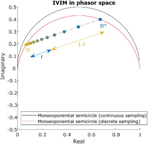
Separating the decay signal from diffusion‐weighted scans into two or more components can be challenging. The phasor technique is well established in the field of optical microscopy for visualization and separation of fluorescent dyes with different lifetimes. The use of the phasor technique for separation of diffusion‐weighted decay signals was recently proposed. In this study, we investigate the added value of this technique for fitting decay models and visualization of decay rates. Phasor visualization was performed in five glioblastoma patients. Using simulations, the influence of incorrect diffusivity values and of the number of b‐values on fitting a three‐component model with fixed diffusivities (dubbed “unmixing”) was investigated for both a phasor‐based fit and a linear least squares (LLS) fit. Phasor‐based intravoxel incoherent motion (IVIM) fitting was compared with nonlinear least squares (NLLS) and segmented fitting (SF) methods in terms of accuracy and precision. The distributions of the parameter estimates of simulated data were compared with those obtained in a healthy volunteer. In the phasor visualizations of two glioblastoma patients, a cluster of points was observed that was not seen in healthy volunteers. The identified cluster roughly corresponded to the enhanced edge region of the tumor of two glioblastoma patients visible on fluid‐attenuated inversion recovery (FLAIR) images. For fitting decay models the usefulness of the phasor transform is less pronounced, but the additional knowledge gained from the geometrical configuration of phasor space can aid fitting routines. This has led to slightly improved fitting results for the IVIM model: phasor‐based fitting yielded parameter maps with higher precision than the NLLS and SF methods for parameters f and D (interquartile range [IQR] for f: NLLS 27, SF 12, phasor 5.7%; IQR for D: NLLS 0.28, SF 0.18, phasor 0.10 μm2/s). For unmixing, LLS fitting slightly but consistently outperformed phasor‐based fitting in all of the tested scenarios.
中文翻译:

使用相量变换解开扩散信号。
将衰减信号从扩散加权扫描分离成两个或多个分量可能具有挑战性。相量技术在光学显微镜领域已经很好地建立起来,用于可视化和分离具有不同寿命的荧光染料。最近提出了使用相量技术来分离扩散加权衰减信号。在这项研究中,我们研究了这种技术在拟合衰减模型和衰减率可视化方面的附加价值。在五名胶质母细胞瘤患者中进行了相量可视化。使用模拟,针对基于相量的拟合和线性最小二乘法 (LLS) 研究了不正确的扩散率值和 b 值数量对拟合具有固定扩散率的三分量模型(称为“分离”)的影响合身。将基于相量的体素内非相干运动 (IVIM) 拟合与非线性最小二乘法 (NLLS) 和分段拟合 (SF) 方法在准确性和精度方面进行了比较。将模拟数据的参数估计分布与在健康志愿者中获得的分布进行比较。在两名胶质母细胞瘤患者的相量可视化中,观察到一组在健康志愿者中没有看到的点。识别出的簇大致对应于在流体衰减反转恢复 (FLAIR) 图像上可见的两名胶质母细胞瘤患者肿瘤的增强边缘区域。对于拟合衰减模型,相量变换的用处不太明显,但从相量空间的几何配置中获得的额外知识可以帮助拟合例程。2 / 秒)。对于解混,LLS 拟合在所有测试场景中略微但始终优于基于相量的拟合。
更新日期:2020-07-23
中文翻译:
使用相量变换解开扩散信号。
将衰减信号从扩散加权扫描分离成两个或多个分量可能具有挑战性。相量技术在光学显微镜领域已经很好地建立起来,用于可视化和分离具有不同寿命的荧光染料。最近提出了使用相量技术来分离扩散加权衰减信号。在这项研究中,我们研究了这种技术在拟合衰减模型和衰减率可视化方面的附加价值。在五名胶质母细胞瘤患者中进行了相量可视化。使用模拟,针对基于相量的拟合和线性最小二乘法 (LLS) 研究了不正确的扩散率值和 b 值数量对拟合具有固定扩散率的三分量模型(称为“分离”)的影响合身。将基于相量的体素内非相干运动 (IVIM) 拟合与非线性最小二乘法 (NLLS) 和分段拟合 (SF) 方法在准确性和精度方面进行了比较。将模拟数据的参数估计分布与在健康志愿者中获得的分布进行比较。在两名胶质母细胞瘤患者的相量可视化中,观察到一组在健康志愿者中没有看到的点。识别出的簇大致对应于在流体衰减反转恢复 (FLAIR) 图像上可见的两名胶质母细胞瘤患者肿瘤的增强边缘区域。对于拟合衰减模型,相量变换的用处不太明显,但从相量空间的几何配置中获得的额外知识可以帮助拟合例程。2 / 秒)。对于解混,LLS 拟合在所有测试场景中略微但始终优于基于相量的拟合。


 京公网安备 11010802027423号