当前位置:
X-MOL 学术
›
NMR Biomed.
›
论文详情
Our official English website, www.x-mol.net, welcomes your
feedback! (Note: you will need to create a separate account there.)
Relaxometry and quantification in sodium MRI of cerebral gliomas: A FET-PET and MRI small-scale study.
NMR in Biomedicine ( IF 2.7 ) Pub Date : 2020-07-21 , DOI: 10.1002/nbm.4361 Wieland A Worthoff 1 , Aliaksandra Shymanskaya 2 , Johannes Lindemeyer 1 , Karl-Josef Langen 1, 3 , N Jon Shah 1, 2, 4
NMR in Biomedicine ( IF 2.7 ) Pub Date : 2020-07-21 , DOI: 10.1002/nbm.4361 Wieland A Worthoff 1 , Aliaksandra Shymanskaya 2 , Johannes Lindemeyer 1 , Karl-Josef Langen 1, 3 , N Jon Shah 1, 2, 4
Affiliation
|
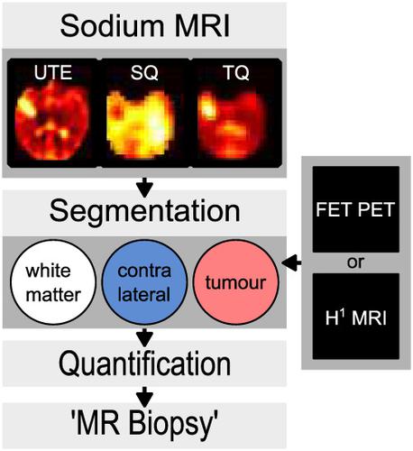
Sodium MRI is a promising method for assessing the metabolic properties of brain tumours. In a recent study, a strong relationship between semi‐quantitative abnormalities in sodium MRI and the mutational status of the isocitrate dehydrogenase enzyme (IDH) with untreated cerebral gliomas was observed. Here, sodium relaxometry in brain tumour tissue was investigated in relation to molecular markers in order to reveal quantitative sodium tissue parameters and the differences between healthy tissue and brain tumour. The previous semi‐quantitative approach is extended by use of suitable relaxometry methods accompanied by numerical simulation to achieve detailed quantitative analysis of intra‐ and extracellular sodium concentration using an enhanced SISTINA sequence at 4 T. Using optimised techniques, biexponential sodium relaxation times in tumour (T*2f, T*2s) and in healthy contralateral brain tissue (T*2f,CL, T*2s,CL) were estimated in 10 patients, along with intracellular sodium molar fractions (χ, χCL), volume fractions (η, ηCL) and concentrations (ρin, ρin,CL). The total sodium tissue concentrations (ρT, ρT,CL) were also estimated. The ratios T*2f/T*2f,CL (P = .05), η/ηCL (P = .02) and χ/χCL (P = .02) were significantly lower in IDH mutated than in IDH wildtype gliomas (n = 4 and n = 5 patients, respectively). The Wilcoxon rank‐sum test was used to compare sodium MRI parameters in patients with and without IDH mutation. Thus, quantitative analysis of relaxation rates, intra‐ and extracellular sodium concentrations, intracellular molar and volume fractions based on enhanced SISTINA confirmed a relationship between abnormalities in sodium parameters and the IDH mutational status in cerebral gliomas, hence catering for the potential to provide further insights into the status of the disease.
中文翻译:

脑胶质瘤钠 MRI 的弛豫测量和量化:FET-PET 和 MRI 小规模研究。
钠 MRI 是一种很有前景的评估脑肿瘤代谢特性的方法。在最近的一项研究中,观察到钠 MRI 的半定量异常与未治疗的脑胶质瘤中异柠檬酸脱氢酶 (IDH) 的突变状态之间存在密切关系。在这里,为了揭示定量的钠组织参数以及健康组织和脑肿瘤之间的差异,研究了脑肿瘤组织中的钠弛豫测量与分子标记的关系。先前的半定量方法通过使用合适的弛豫方法以及数值模拟进行扩展,以使用增强的 SISTINA 序列在 4 T 下实现细胞内和细胞外钠浓度的详细定量分析。 使用优化技术,双指数钠弛豫时间在肿瘤中(T* 2f , T* 2s ) 和健康对侧脑组织 ( T* 2f,CL , T* 2s,CL ) 估计了 10 名患者,以及细胞内钠摩尔分数 (χ, χ CL )、体积分数 (η , η CL ) 和浓度 (ρ in , ρ in,CL )。还估计了总钠组织浓度 (ρ T , ρ T, CL )。比率T * 2f / T * 2f,CL ( P = .05), η/η CL ( P = .02) 和 χ/χIDH 突变的CL ( P = .02) 显着低于 IDH 野生型神经胶质瘤(分别为n = 4 和n = 5 名患者)。Wilcoxon 秩和检验用于比较有和没有 IDH 突变患者的钠 MRI 参数。因此,基于增强的 SISTINA 对舒张率、细胞内和细胞外钠浓度、细胞内摩尔和体积分数的定量分析证实了钠参数异常与脑胶质瘤中 IDH 突变状态之间的关系,因此有可能提供进一步的见解进入疾病状态。
更新日期:2020-09-03
中文翻译:
脑胶质瘤钠 MRI 的弛豫测量和量化:FET-PET 和 MRI 小规模研究。
钠 MRI 是一种很有前景的评估脑肿瘤代谢特性的方法。在最近的一项研究中,观察到钠 MRI 的半定量异常与未治疗的脑胶质瘤中异柠檬酸脱氢酶 (IDH) 的突变状态之间存在密切关系。在这里,为了揭示定量的钠组织参数以及健康组织和脑肿瘤之间的差异,研究了脑肿瘤组织中的钠弛豫测量与分子标记的关系。先前的半定量方法通过使用合适的弛豫方法以及数值模拟进行扩展,以使用增强的 SISTINA 序列在 4 T 下实现细胞内和细胞外钠浓度的详细定量分析。 使用优化技术,双指数钠弛豫时间在肿瘤中(T* 2f , T* 2s ) 和健康对侧脑组织 ( T* 2f,CL , T* 2s,CL ) 估计了 10 名患者,以及细胞内钠摩尔分数 (χ, χ CL )、体积分数 (η , η CL ) 和浓度 (ρ in , ρ in,CL )。还估计了总钠组织浓度 (ρ T , ρ T, CL )。比率T * 2f / T * 2f,CL ( P = .05), η/η CL ( P = .02) 和 χ/χIDH 突变的CL ( P = .02) 显着低于 IDH 野生型神经胶质瘤(分别为n = 4 和n = 5 名患者)。Wilcoxon 秩和检验用于比较有和没有 IDH 突变患者的钠 MRI 参数。因此,基于增强的 SISTINA 对舒张率、细胞内和细胞外钠浓度、细胞内摩尔和体积分数的定量分析证实了钠参数异常与脑胶质瘤中 IDH 突变状态之间的关系,因此有可能提供进一步的见解进入疾病状态。


 京公网安备 11010802027423号