当前位置:
X-MOL 学术
›
Cryst. Res. Technol.
›
论文详情
Our official English website, www.x-mol.net, welcomes your feedback! (Note: you will need to create a separate account there.)
X‐Ray Topography—More than Nice Pictures
Crystal Research and Technology ( IF 1.5 ) Pub Date : 2020-07-22 , DOI: 10.1002/crat.202000012 Andreas N. Danilewsky 1
Crystal Research and Technology ( IF 1.5 ) Pub Date : 2020-07-22 , DOI: 10.1002/crat.202000012 Andreas N. Danilewsky 1
Affiliation
|
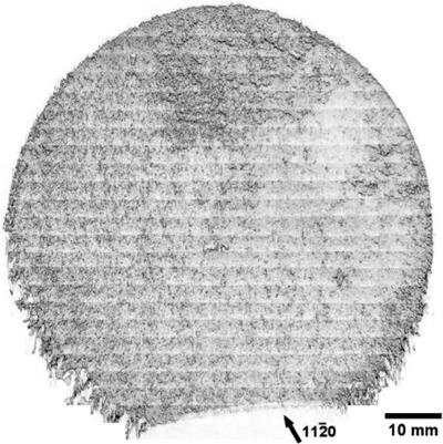
X‐ray topography—a well‐known diffraction imaging method—is widely used for the characterization of extended crystal defects like dislocations. Herein, the progress toward the quantification besides the number and nature of dislocations is given. The diffracted images include additional information about tilt and strain, which can be measured in real‐time, thanks to the improved digital detection systems. This allows not only the fast mapping of huge samples like 450 mm diameter Si wafers, the in situ observation of the dislocation, or crack dynamics at high temperatures, but also the stress analysis in electronic devices in operando. In addition to typical white X‐ray beam methods like stacks of section topographs, the monochromatic diffraction laminography allows a 3D representation of the dislocation arrays. The merits and limitations of X‐ray topography are demonstrated by semiconductor materials as an example.
中文翻译:

X射线地形图-不仅仅是尼斯图片
X射线形貌是一种众所周知的衍射成像方法,被广泛用于表征位错等扩展的晶体缺陷。在此,除了位错的数量和性质之外,还给出了向量化的进展。由于改进了数字检测系统,衍射图像还包含有关倾斜和应变的附加信息,这些信息可以实时测量。这不仅可以快速绘制大型样品(如450毫米直径的硅晶圆),可以对位错进行原位观察,也可以在高温下观察裂纹动态,还可以进行电子设备中的应力分析。除了典型的白色X射线束方法(如截面形貌图的叠加)外,单色衍射层照相术还可以对位错阵列进行3D表示。
更新日期:2020-09-09
中文翻译:
X射线地形图-不仅仅是尼斯图片
X射线形貌是一种众所周知的衍射成像方法,被广泛用于表征位错等扩展的晶体缺陷。在此,除了位错的数量和性质之外,还给出了向量化的进展。由于改进了数字检测系统,衍射图像还包含有关倾斜和应变的附加信息,这些信息可以实时测量。这不仅可以快速绘制大型样品(如450毫米直径的硅晶圆),可以对位错进行原位观察,也可以在高温下观察裂纹动态,还可以进行电子设备中的应力分析。除了典型的白色X射线束方法(如截面形貌图的叠加)外,单色衍射层照相术还可以对位错阵列进行3D表示。


 京公网安备 11010802027423号