当前位置:
X-MOL 学术
›
Microsc. Res. Tech.
›
论文详情
Our official English website, www.x-mol.net, welcomes your
feedback! (Note: you will need to create a separate account there.)
Measurement of erosion depth using microcomputed tomography and light microscopy.
Microscopy Research and Technique ( IF 2.0 ) Pub Date : 2020-07-18 , DOI: 10.1002/jemt.23537 Gina Delia Roque-Torres 1 , So Ran Kwon 1 , Udochukwu Oyoyo 1 , Yiming Li 1
Microscopy Research and Technique ( IF 2.0 ) Pub Date : 2020-07-18 , DOI: 10.1002/jemt.23537 Gina Delia Roque-Torres 1 , So Ran Kwon 1 , Udochukwu Oyoyo 1 , Yiming Li 1
Affiliation
|
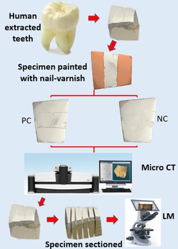
Tooth‐erosion is the surface loss of dental hard tissue mostly associated with an acid attack. The aim was to compare dentin and enamel erosion depth measurements using micro‐computed tomography (microCT) and light microscopy (LM). Enamel/dentin blocks were prepared from caries‐free human molar‐teeth (N = 12). Teeth were sectioned to a rectangular shape of 4 × 4 × 6 mm. Specimens were treated with water (NC) or 1.0% citric‐acid solution (PC). After treatment, specimens were scanned with micro‐computed tomography. On completion, specimens were sectioned and observed under a light‐microscope. Lesion depth was observed with 10× magnification and images transferred to Simpleware software. Vertical distance from lesion surface to bottom was measured. Pearson correlation test was used to evaluate correlation and Wilcoxon Signed Rank test to evaluate differences in the two‐analysis methods. Mean enamel erosion depth was 0.63 and 38.38 μm (microCT) and 0.54 and 39.43 μm (LM) for NC and PC, respectively. Dentin erosion depth was 0.72 and 48.05 μm (microCT) and 0.56 and 49.92 μm (LM) for NC and PC, respectively. There was a significant correlation between the two‐analysis methods (r = 0.998; p < .001). No statistically significant difference in results were obtained when microCT and LM were compared (p = .584). This results obtained from the current study suggested that erosion depth measurements made using microCT and LM yielded comparable results. The microCT method is preferred if the conservation of specimens is desired.
中文翻译:

使用显微计算机断层扫描和光学显微镜测量侵蚀深度。
牙齿侵蚀是牙齿硬组织的表面损失,主要与酸侵蚀有关。目的是使用显微计算机断层扫描 (microCT) 和光学显微镜 (LM) 来比较牙本质和牙釉质侵蚀深度测量值。牙釉质/牙本质块由无龋人臼齿制备(N= 12)。牙齿被切成4×4×6mm的矩形。样品用水 (NC) 或 1.0% 柠檬酸溶液 (PC) 处理。治疗后,用显微计算机断层扫描对标本进行扫描。完成后,将样品切片并在光学显微镜下观察。用 10 倍放大率观察病变深度,并将图像传输到 Simpleware 软件。测量从病变表面到底部的垂直距离。Pearson 相关性检验用于评估相关性,Wilcoxon Signed Rank 检验用于评估两种分析方法的差异。NC 和 PC 的平均牙釉质侵蚀深度分别为 0.63 和 38.38 μm (microCT) 以及 0.54 和 39.43 μm (LM)。NC 和 PC 的牙本质侵蚀深度分别为 0.72 和 48.05 μm (microCT) 以及 0.56 和 49.92 μm (LM)。r = 0.998;p < .001)。当比较 microCT 和 LM 时,结果没有统计学上的显着差异 ( p = .584)。从当前研究中获得的这一结果表明,使用 microCT 和 LM 进行的侵蚀深度测量产生了可比较的结果。如果需要保存标本,则首选 microCT 方法。
更新日期:2020-07-18
中文翻译:
使用显微计算机断层扫描和光学显微镜测量侵蚀深度。
牙齿侵蚀是牙齿硬组织的表面损失,主要与酸侵蚀有关。目的是使用显微计算机断层扫描 (microCT) 和光学显微镜 (LM) 来比较牙本质和牙釉质侵蚀深度测量值。牙釉质/牙本质块由无龋人臼齿制备(N= 12)。牙齿被切成4×4×6mm的矩形。样品用水 (NC) 或 1.0% 柠檬酸溶液 (PC) 处理。治疗后,用显微计算机断层扫描对标本进行扫描。完成后,将样品切片并在光学显微镜下观察。用 10 倍放大率观察病变深度,并将图像传输到 Simpleware 软件。测量从病变表面到底部的垂直距离。Pearson 相关性检验用于评估相关性,Wilcoxon Signed Rank 检验用于评估两种分析方法的差异。NC 和 PC 的平均牙釉质侵蚀深度分别为 0.63 和 38.38 μm (microCT) 以及 0.54 和 39.43 μm (LM)。NC 和 PC 的牙本质侵蚀深度分别为 0.72 和 48.05 μm (microCT) 以及 0.56 和 49.92 μm (LM)。r = 0.998;p < .001)。当比较 microCT 和 LM 时,结果没有统计学上的显着差异 ( p = .584)。从当前研究中获得的这一结果表明,使用 microCT 和 LM 进行的侵蚀深度测量产生了可比较的结果。如果需要保存标本,则首选 microCT 方法。


 京公网安备 11010802027423号