当前位置:
X-MOL 学术
›
Microsc. Res. Tech.
›
论文详情
Our official English website, www.x-mol.net, welcomes your
feedback! (Note: you will need to create a separate account there.)
Holotomographic microscopy: A new approach to detect apoptotic cell features.
Microscopy Research and Technique ( IF 2.0 ) Pub Date : 2020-07-18 , DOI: 10.1002/jemt.23539 Sara Salucci 1 , Michela Battistelli 1 , Sabrina Burattini 1 , Francesca Sbrana 2 , Elisabetta Falcieri 1
Microscopy Research and Technique ( IF 2.0 ) Pub Date : 2020-07-18 , DOI: 10.1002/jemt.23539 Sara Salucci 1 , Michela Battistelli 1 , Sabrina Burattini 1 , Francesca Sbrana 2 , Elisabetta Falcieri 1
Affiliation
|
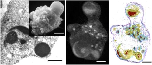
Holotomographic (HT) microscopy, combines two techniques, holography and tomography, and, in this way, it allows to quantitatively and noninvasively investigate cells and thin tissue slices, by obtaining three‐dimensional (3D) images and by monitoring inner morphological changes. HT has indeed two significant advantages: it is label‐free and low‐energy light passes through the specimen with minimal perturbation. Using quantitative phase imaging with optical diffraction tomography, it can produce 3D images by measuring the refraction index (RI). Therefore, based on RI values, HT can provide structural and chemical cell information, such as dry mass values, morphological changes, or cellular membrane dynamics. In this study, suspended and adherent culture cells have been processed for HT analyses. Some of them have been treated with known apoptotic drugs or pro‐oxidant agents and cell response has been investigated both by conventional microscopic approaches and by HT. The ultrastructural and fluorescence images have been compared to those obtained by HT and their congruence has been discussed, with particular attention to apoptotic cell death and on correlated plasma membrane changes. HT appears a valid approach to further characterize well‐known apoptotic features such as cell blebbing, chromatin condensation, micronuclei, and apoptotic bodies. Taken together, our data demonstrate that HT appears suitable to highlight suspended or adherent cell behavior under different conditions. In particular, this technique appears an important new tool to distinguish healthy cells from the apoptotic ones, as well as to monitor outer and inner cell changes in a rapid way and with a noninvasive, label‐free, approach.
中文翻译:

全息显微镜:一种检测凋亡细胞特征的新方法。
全息 (HT) 显微镜结合了全息和断层扫描两种技术,通过这种方式,它可以通过获取三维 (3D) 图像和监测内部形态变化,以定量和无创的方式研究细胞和薄组织切片。HT 确实有两个显着的优点:它是无标记的,低能量的光以最小的扰动穿过样品。使用带有光学衍射断层扫描的定量相位成像,它可以通过测量折射率 (RI) 产生 3D 图像。因此,基于 RI 值,HT 可以提供结构和化学细胞信息,例如干重值、形态变化或细胞膜动力学。在这项研究中,悬浮和贴壁培养细胞已被处理用于 HT 分析。其中一些已用已知的凋亡药物或促氧化剂处理,并且已通过常规显微镜方法和 HT 研究了细胞反应。将超微结构和荧光图像与 HT 获得的图像进行了比较,并讨论了它们的一致性,特别注意凋亡细胞死亡和相关的质膜变化。HT 似乎是进一步表征众所周知的凋亡特征的有效方法,例如细胞起泡、染色质凝聚、微核和凋亡小体。总之,我们的数据表明 HT 似乎适合突出不同条件下的悬浮或贴壁细胞行为。特别是,这项技术似乎是区分健康细胞和凋亡细胞的重要新工具,
更新日期:2020-07-18
中文翻译:
全息显微镜:一种检测凋亡细胞特征的新方法。
全息 (HT) 显微镜结合了全息和断层扫描两种技术,通过这种方式,它可以通过获取三维 (3D) 图像和监测内部形态变化,以定量和无创的方式研究细胞和薄组织切片。HT 确实有两个显着的优点:它是无标记的,低能量的光以最小的扰动穿过样品。使用带有光学衍射断层扫描的定量相位成像,它可以通过测量折射率 (RI) 产生 3D 图像。因此,基于 RI 值,HT 可以提供结构和化学细胞信息,例如干重值、形态变化或细胞膜动力学。在这项研究中,悬浮和贴壁培养细胞已被处理用于 HT 分析。其中一些已用已知的凋亡药物或促氧化剂处理,并且已通过常规显微镜方法和 HT 研究了细胞反应。将超微结构和荧光图像与 HT 获得的图像进行了比较,并讨论了它们的一致性,特别注意凋亡细胞死亡和相关的质膜变化。HT 似乎是进一步表征众所周知的凋亡特征的有效方法,例如细胞起泡、染色质凝聚、微核和凋亡小体。总之,我们的数据表明 HT 似乎适合突出不同条件下的悬浮或贴壁细胞行为。特别是,这项技术似乎是区分健康细胞和凋亡细胞的重要新工具,


 京公网安备 11010802027423号