当前位置:
X-MOL 学术
›
NMR Biomed.
›
论文详情
Our official English website, www.x-mol.net, welcomes your
feedback! (Note: you will need to create a separate account there.)
Fast quantitative three-dimensional ultrashort echo time (UTE) Cones magnetic resonance imaging of major tissues in the knee joint using extended sprial sampling.
NMR in Biomedicine ( IF 2.7 ) Pub Date : 2020-07-15 , DOI: 10.1002/nbm.4376 Lidi Wan 1, 2 , Yajun Ma 2 , Jiawei Yang 1 , Saeed Jerban 2 , Adam C Searleman 2 , Michael Carl 3 , Nicole Le 4 , Eric Y Chang 2, 4 , Guangyu Tang 1 , Jiang Du 2
NMR in Biomedicine ( IF 2.7 ) Pub Date : 2020-07-15 , DOI: 10.1002/nbm.4376 Lidi Wan 1, 2 , Yajun Ma 2 , Jiawei Yang 1 , Saeed Jerban 2 , Adam C Searleman 2 , Michael Carl 3 , Nicole Le 4 , Eric Y Chang 2, 4 , Guangyu Tang 1 , Jiang Du 2
Affiliation
|
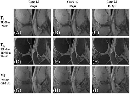
The purpose of this study is to investigate the effect of extending the spiral sampling window on quantitative 3D ultrashort echo time (UTE) Cones imaging of major knee joint tissues including articular cartilage, menisci, tendons and ligaments at 3 T. Nine cadaveric human whole knee specimens were imaged on a 3 T clinical MRI scanner. A series of quantitative 3D UTE Cones imaging biomarkers including T2*, T1, adiabatic T1ρ, magnetization transfer ratio (MTR) and macromolecular fraction (MMF) were estimated using spiral sampling trajectories with various durations. Errors in UTE MRI biomarkers as a function of sampling time were evaluated using a nonstretched spiral trajectory as a reference standard. No significant differences were observed by increasing the spiral sampling window from 1116 to 2232 μs in the calculated T2*, T1, adiabatic T1ρ, MTR and MMF, as all P‐values were over .05 as assessed by ANOVA with two‐sided Dunnett's test. Although extending the sampling window results in signal loss for short T2 components, there was limited effect on the calculated quantitative biomarkers, with error percentages typically smaller than 5% in all the evaluated tissues. The total scan time can be reduced by up to 54% with quantification errors of less than 5% in any evaluated major tissue in the knee joint, suggesting that 3D UTE Cones MRI techniques can be greatly accelerated by using a longer spiral sampling window without causing additional quantitative bias.
中文翻译:

使用扩展螺旋采样对膝关节主要组织进行快速定量三维超短回波时间 (UTE) 锥体磁共振成像。
本研究的目的是研究扩展螺旋采样窗口对主要膝关节组织(包括关节软骨、半月板、肌腱和韧带)的定量 3D 超短回波时间 (UTE) 锥体成像的影响。 9 具尸体人体全膝标本在 3 T 临床 MRI 扫描仪上成像。一系列定量 3D UTE Cones 成像生物标志物,包括 T 2 *、T 1、绝热 T 1ρ,磁化转移比(MTR)和大分子分数(MMF)使用具有不同持续时间的螺旋采样轨迹估计。使用未拉伸的螺旋轨迹作为参考标准评估作为采样时间函数的 UTE MRI 生物标志物的误差。在计算的 T 2 *、T 1、绝热 T 1ρ、MTR 和 MMF 中,通过将螺旋采样窗口从 1116 μs 增加到 2232 μs 没有观察到显着差异,因为所有P 值都超过 0.05,如通过 ANOVA 评估的两个支持 Dunnett 检验。虽然延长采样窗口会导致短 T 2 的信号丢失成分,对计算的定量生物标志物的影响有限,所有评估组织的误差百分比通常小于 5%。总扫描时间最多可减少 54%,膝关节中任何评估的主要组织的量化误差小于 5%,这表明 3D UTE Cones MRI 技术可以通过使用更长的螺旋采样窗口而大大加速,而不会导致额外的定量偏差。
更新日期:2020-09-03
中文翻译:
使用扩展螺旋采样对膝关节主要组织进行快速定量三维超短回波时间 (UTE) 锥体磁共振成像。
本研究的目的是研究扩展螺旋采样窗口对主要膝关节组织(包括关节软骨、半月板、肌腱和韧带)的定量 3D 超短回波时间 (UTE) 锥体成像的影响。 9 具尸体人体全膝标本在 3 T 临床 MRI 扫描仪上成像。一系列定量 3D UTE Cones 成像生物标志物,包括 T 2 *、T 1、绝热 T 1ρ,磁化转移比(MTR)和大分子分数(MMF)使用具有不同持续时间的螺旋采样轨迹估计。使用未拉伸的螺旋轨迹作为参考标准评估作为采样时间函数的 UTE MRI 生物标志物的误差。在计算的 T 2 *、T 1、绝热 T 1ρ、MTR 和 MMF 中,通过将螺旋采样窗口从 1116 μs 增加到 2232 μs 没有观察到显着差异,因为所有P 值都超过 0.05,如通过 ANOVA 评估的两个支持 Dunnett 检验。虽然延长采样窗口会导致短 T 2 的信号丢失成分,对计算的定量生物标志物的影响有限,所有评估组织的误差百分比通常小于 5%。总扫描时间最多可减少 54%,膝关节中任何评估的主要组织的量化误差小于 5%,这表明 3D UTE Cones MRI 技术可以通过使用更长的螺旋采样窗口而大大加速,而不会导致额外的定量偏差。


 京公网安备 11010802027423号