当前位置:
X-MOL 学术
›
Photochem. Photobiol. Sci.
›
论文详情
Our official English website, www.x-mol.net, welcomes your
feedback! (Note: you will need to create a separate account there.)
In flow metal-enhanced fluorescence for biolabelling and biodetection.
Photochemical & Photobiological Sciences ( IF 2.7 ) Pub Date : 2020-07-14 , DOI: 10.1039/d0pp00145g Daniela Gontero 1 , Alicia V Veglia 2 , A Guillermo Bracamonte 2, 3
Photochemical & Photobiological Sciences ( IF 2.7 ) Pub Date : 2020-07-14 , DOI: 10.1039/d0pp00145g Daniela Gontero 1 , Alicia V Veglia 2 , A Guillermo Bracamonte 2, 3
Affiliation
|
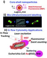
Escherichia coli bacteria were determined by in flow cytometry with laser excitation and fluorescence detection applying ultraluminescent core–shell nanoparticles based on Metal Enhanced Fluorescence (MEF). Core–shell nanoparticles consisted of a 40 nm core modified with a silica spacer grafted with Rhodamine B (RhB). The electromagnetic field in the near field of the core surface enhanced the fluorescence of RhB by plasmonic and fluorophore coupling. The hydrophilic silica spacer allowed the non-covalent interaction with the polar E. coli surface and thus ultraluminescent bacteria biolabelling was developed. Clearly, well defined and bright bacteria imaging was recorded by Laser Fluorescence Microscopy based on the non-covalent deposition of the ultraluminescent nano-emitters. Using these nano-labellers, it was possible to detect labelled E. coli by in flow cytometry. Higher values of Side-scattered light (SSC) and Forward-scattered light (FSC), and number of fluorescent event detections, were observed for labelled bacteria compared to those non-labelled. The sensitivity of the methodology was evaluated by varying bacteria concentration and acceptable analytical figures of merit were determined. Applying this methodology we could quantify E. coli from a synthetic real sample of fortified water. Similar results were obtained by bacteria counting with Laser Fluorescence Microscopy and with a cell-bacteria counter.
中文翻译:

在流动中,金属增强的荧光用于生物标记和生物检测。
大肠杆菌是通过流式细胞术,激光激发和基于金属增强荧光(MEF)的超发光核-壳纳米粒子的荧光检测而确定的。核-壳纳米颗粒由40 nm核组成,该核被嫁接了若丹明B(RhB)的二氧化硅间隔物修饰。核心表面近场中的电磁场通过等离激元和荧光团偶联增强了RhB的荧光。亲水性二氧化硅间隔基允许与极性大肠杆菌进行非共价相互作用表面,因此开发了超发光细菌生物标记。显然,基于超发光纳米发射体的非共价沉积,通过激光荧光显微镜记录了清晰清晰的细菌成像。使用这些纳米标记物,可以通过流式细胞术检测标记的大肠杆菌。与未标记细菌相比,标记细菌观察到了更高的侧向散射光(SSC)和前向散射光(FSC)值,以及荧光事件检测次数。通过改变细菌浓度来评估该方法的敏感性,并确定可接受的品质因数。应用这种方法,我们可以量化大肠杆菌来自强化水的合成真实样本。通过使用激光荧光显微镜和细胞细菌计数器进行细菌计数获得了相似的结果。
更新日期:2020-09-16
中文翻译:
在流动中,金属增强的荧光用于生物标记和生物检测。
大肠杆菌是通过流式细胞术,激光激发和基于金属增强荧光(MEF)的超发光核-壳纳米粒子的荧光检测而确定的。核-壳纳米颗粒由40 nm核组成,该核被嫁接了若丹明B(RhB)的二氧化硅间隔物修饰。核心表面近场中的电磁场通过等离激元和荧光团偶联增强了RhB的荧光。亲水性二氧化硅间隔基允许与极性大肠杆菌进行非共价相互作用表面,因此开发了超发光细菌生物标记。显然,基于超发光纳米发射体的非共价沉积,通过激光荧光显微镜记录了清晰清晰的细菌成像。使用这些纳米标记物,可以通过流式细胞术检测标记的大肠杆菌。与未标记细菌相比,标记细菌观察到了更高的侧向散射光(SSC)和前向散射光(FSC)值,以及荧光事件检测次数。通过改变细菌浓度来评估该方法的敏感性,并确定可接受的品质因数。应用这种方法,我们可以量化大肠杆菌来自强化水的合成真实样本。通过使用激光荧光显微镜和细胞细菌计数器进行细菌计数获得了相似的结果。


 京公网安备 11010802027423号