当前位置:
X-MOL 学术
›
Prog. Nat. Sci. Mater. Int.
›
论文详情
Our official English website, www.x-mol.net, welcomes your
feedback! (Note: you will need to create a separate account there.)
Nano to micro size transition of hydroxyapatite in porcine bone during heat treatment with low heating rates
Progress in Natural Science: Materials International ( IF 4.8 ) Pub Date : 2020-08-01 , DOI: 10.1016/j.pnsc.2020.06.005 Angélica M. Castillo-Paz , Sandra M. Londoño-Restrepo , Liliana Tirado-Mejía , M.A. Mondragón , Mario E. Rodríguez-García
Progress in Natural Science: Materials International ( IF 4.8 ) Pub Date : 2020-08-01 , DOI: 10.1016/j.pnsc.2020.06.005 Angélica M. Castillo-Paz , Sandra M. Londoño-Restrepo , Liliana Tirado-Mejía , M.A. Mondragón , Mario E. Rodríguez-García
|
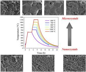
Abstract This work focuses on the changes of raw hydroxyapatite obtained from porcine bones during heat treatment with heating rates of 2.5 and 5.0 °C/min between 600 °C and 1100 °C. High-Resolution transmission electron microscopy (HR-TEM) images showed that raw hydroxyapatite was formed by high-ordered nanometric platelet crystals with a length of around 18 nm and width around 7 nm. High-Resolution scanning electron microscopy (HR-SEM) images evidenced the coalescence phenomena during heating giving. It was found that the growth of the crystals and the transition from nano to a micro-size occurred at around 700 °C. And the changes in the full width at half maximum (FWHM) for the characteristic (002) plane of the X-ray diffraction patterns and the FWHM of the characteristic Raman band at 960 cm-1 for ν1 PO43- exhibited the transition from nano to a micro size, that was confirmed by HR-SEM images and the thermogravimetric (TG) analysis. The porosity of the BIO-HAp changes from intra nanometric to intercrystalline.
中文翻译:

低加热速率热处理过程中猪骨中羟基磷灰石的纳米到微米尺寸转变
摘要 本工作重点研究了从猪骨中获得的原料羟基磷灰石在 600°C 和 1100°C 之间以 2.5 和 5.0°C/min 的加热速率进行热处理的变化。高分辨率透射电子显微镜 (HR-TEM) 图像显示,原始羟基磷灰石由长约 18 nm、宽约 7 nm 的高有序纳米片状晶体形成。高分辨率扫描电子显微镜 (HR-SEM) 图像证明了加热过程中的聚结现象。发现晶体的生长和从纳米尺寸到微米尺寸的转变发生在 700 °C 左右。X 射线衍射图的特征 (002) 面的半峰全宽 (FWHM) 和 ν1 PO43- 的特征拉曼谱带在 960 cm-1 处的 FWHM 的变化表现出从纳米到微小尺寸,这通过 HR-SEM 图像和热重 (TG) 分析得到证实。BIO-HAp 的孔隙率从纳米内变为晶间。
更新日期:2020-08-01
中文翻译:
低加热速率热处理过程中猪骨中羟基磷灰石的纳米到微米尺寸转变
摘要 本工作重点研究了从猪骨中获得的原料羟基磷灰石在 600°C 和 1100°C 之间以 2.5 和 5.0°C/min 的加热速率进行热处理的变化。高分辨率透射电子显微镜 (HR-TEM) 图像显示,原始羟基磷灰石由长约 18 nm、宽约 7 nm 的高有序纳米片状晶体形成。高分辨率扫描电子显微镜 (HR-SEM) 图像证明了加热过程中的聚结现象。发现晶体的生长和从纳米尺寸到微米尺寸的转变发生在 700 °C 左右。X 射线衍射图的特征 (002) 面的半峰全宽 (FWHM) 和 ν1 PO43- 的特征拉曼谱带在 960 cm-1 处的 FWHM 的变化表现出从纳米到微小尺寸,这通过 HR-SEM 图像和热重 (TG) 分析得到证实。BIO-HAp 的孔隙率从纳米内变为晶间。


 京公网安备 11010802027423号