当前位置:
X-MOL 学术
›
J. Cell. Physiol.
›
论文详情
Our official English website, www.x-mol.net, welcomes your
feedback! (Note: you will need to create a separate account there.)
Increased cellular senescence in the murine and human stenotic kidney: Effect of mesenchymal stem cells.
Journal of Cellular Physiology ( IF 4.5 ) Pub Date : 2020-07-13 , DOI: 10.1002/jcp.29940 Seo Rin Kim 1 , Xiangyu Zou 1 , Hui Tang 1 , Amrutesh S Puranik 1 , Abdelrhman M Abumoawad 1 , Xiang-Yang Zhu 1 , LaTonya J Hickson 1 , Tamara Tchkonia 2 , Stephen C Textor 1 , James L Kirkland 2 , Lilach O Lerman 1
Journal of Cellular Physiology ( IF 4.5 ) Pub Date : 2020-07-13 , DOI: 10.1002/jcp.29940 Seo Rin Kim 1 , Xiangyu Zou 1 , Hui Tang 1 , Amrutesh S Puranik 1 , Abdelrhman M Abumoawad 1 , Xiang-Yang Zhu 1 , LaTonya J Hickson 1 , Tamara Tchkonia 2 , Stephen C Textor 1 , James L Kirkland 2 , Lilach O Lerman 1
Affiliation
|
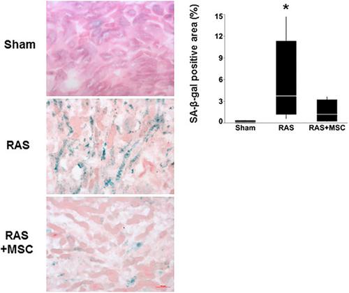
Cell stress may give rise to insuperable growth arrest, which is defined as cellular senescence. Stenotic kidney (STK) ischemia and injury induced by renal artery stenosis (RAS) may be associated with cellular senescence. Mesenchymal stem cells (MSCs) decrease some forms of STK injury, but their ability to reverse senescence in RAS remains unknown. We hypothesized that RAS evokes STK senescence, which would be ameliorated by MSCs. Mice were studied after 4 weeks of RAS, RAS treated with adipose tissue‐derived MSCs 2 weeks earlier, or sham. STK senescence‐associated β‐galactosidase (SA‐β‐Gal) activity was measured. Protein and gene expression was used to assess senescence and the senescence‐associated secretory phenotype (SASP), and staining for renal fibrosis, inflammation, and capillary density. In addition, senescence was assessed as p16+ and p21+ urinary exosomes in patients with renovascular hypertension (RVH) without or 3 months after autologous adipose tissue‐derived MSC delivery, and in healthy volunteers (HV). In RAS mice, STK SA‐β‐Gal activity increased, and senescence and SASP marker expression was markedly elevated. MSCs improved renal function, fibrosis, inflammation, and capillary density, and attenuated SA‐β‐Gal activity, but most senescence and SASP levels remained unchanged. Congruently, in human RVH, p21+ urinary exosomes were elevated compared to HV, and only slightly improved by MSC, whereas p16+ exosomes remained unchanged. Therefore, RAS triggers renal senescence in both mice and human subjects. MSCs decrease renal injury, but only partly mitigate renal senescence. These observations support exploration of targeted senolytic therapy in RAS.
中文翻译:

小鼠和人狭窄肾细胞衰老增加:间充质干细胞的作用。
细胞应激可能导致无法克服的生长停滞,这被定义为细胞衰老。肾动脉狭窄(RAS)引起的狭窄肾(STK)缺血和损伤可能与细胞衰老有关。间充质干细胞 (MSCs) 减少了某些形式的 STK 损伤,但它们逆转 RAS 衰老的能力仍然未知。我们假设 RAS 引起 STK 衰老,这将被 MSC 改善。在 RAS 4 周后对小鼠进行了研究,2 周前用脂肪组织来源的 MSC 治疗 RAS 或假手术。测量了 STK 衰老相关的 β-半乳糖苷酶 (SA-β-Gal) 活性。蛋白质和基因表达用于评估衰老和衰老相关分泌表型 (SASP),以及肾纤维化、炎症和毛细血管密度的染色。此外,衰老被评估为肾血管性高血压 (RVH) 患者和自体脂肪组织来源的 MSC 递送后 3 个月和健康志愿者 (HV) 的p 16+ 和p 21+ 尿外泌体。在 RAS 小鼠中,STK SA-β-Gal 活性增加,衰老和 SASP 标志物表达显着升高。间充质干细胞改善肾功能、纤维化、炎症和毛细血管密度,并减弱 SA-β-Gal 活性,但大多数衰老和 SASP 水平保持不变。一致地,在人类 RVH 中,p与 HV 相比,21+ 尿外泌体升高,MSC 仅略有改善,而 p16+ 外泌体保持不变。因此,RAS 引发小鼠和人类受试者的肾衰老。间充质干细胞减少肾损伤,但仅部分减轻肾衰老。这些观察结果支持在 RAS 中探索靶向抗衰老治疗。
更新日期:2020-07-13
中文翻译:
小鼠和人狭窄肾细胞衰老增加:间充质干细胞的作用。
细胞应激可能导致无法克服的生长停滞,这被定义为细胞衰老。肾动脉狭窄(RAS)引起的狭窄肾(STK)缺血和损伤可能与细胞衰老有关。间充质干细胞 (MSCs) 减少了某些形式的 STK 损伤,但它们逆转 RAS 衰老的能力仍然未知。我们假设 RAS 引起 STK 衰老,这将被 MSC 改善。在 RAS 4 周后对小鼠进行了研究,2 周前用脂肪组织来源的 MSC 治疗 RAS 或假手术。测量了 STK 衰老相关的 β-半乳糖苷酶 (SA-β-Gal) 活性。蛋白质和基因表达用于评估衰老和衰老相关分泌表型 (SASP),以及肾纤维化、炎症和毛细血管密度的染色。此外,衰老被评估为肾血管性高血压 (RVH) 患者和自体脂肪组织来源的 MSC 递送后 3 个月和健康志愿者 (HV) 的p 16+ 和p 21+ 尿外泌体。在 RAS 小鼠中,STK SA-β-Gal 活性增加,衰老和 SASP 标志物表达显着升高。间充质干细胞改善肾功能、纤维化、炎症和毛细血管密度,并减弱 SA-β-Gal 活性,但大多数衰老和 SASP 水平保持不变。一致地,在人类 RVH 中,p与 HV 相比,21+ 尿外泌体升高,MSC 仅略有改善,而 p16+ 外泌体保持不变。因此,RAS 引发小鼠和人类受试者的肾衰老。间充质干细胞减少肾损伤,但仅部分减轻肾衰老。这些观察结果支持在 RAS 中探索靶向抗衰老治疗。


 京公网安备 11010802027423号