Cellular Signalling ( IF 4.8 ) Pub Date : 2020-07-09 , DOI: 10.1016/j.cellsig.2020.109710 Anna Pintér 1 , Zsófia Hevesi 2 , Péter Zahola 3 , Alán Alpár 3 , János Hanics 3
|
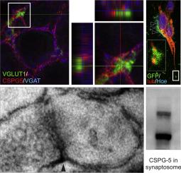
Composition of the brain extracellular matrix changes in time as maturation proceeds. Chondroitin sulfate proteoglycan 5 (CSPG-5), also known as neuroglycan C, has been previously associated to differentiation since it shapes neurite growth and synapse forming. Here, we show that this proteoglycan persists in the postnatal rat brain, and its expression is higher in cortical regions with plastic properties, including hippocampus and the medial prefrontal cortex at the end of the second postnatal week. Progressively accumulating after birth, CSPG-5 typically concentrates around glutamatergic and GABAergic terminals in twelve-week old rat hippocampus. CSPG-5-containing perisynaptic matrix rings often appear at the peripheral margin of perineuronal nets. Electron microscopy and analysis of synaptosomal fraction showed that CSPG-5 accumulates around, and is associated to synapses, respectively. In vitro analyses suggest that neurons, but less so astrocytes, express CSPG-5 in rat primary neocortical cultures, and CSPG-5 produced by transfected neuroblastoma cells appear at endings and contact points of neurites. In human subjects, CSPG-5 expression shifts in brain areas of the default mode network of suicide victims, which may reflect an impact in the pathogenesis of psychiatric diseases or support diagnostic power.
中文翻译:
硫酸软骨素蛋白多糖-5 在成年大鼠皮质中形成突触周围基质组件。
随着成熟的进行,脑细胞外基质的组成随时间而变化。硫酸软骨素蛋白多糖 5 (CSPG-5),也称为神经多糖 C,以前与分化有关,因为它塑造了神经突生长和突触形成。在这里,我们表明这种蛋白多糖在出生后的大鼠大脑中持续存在,并且其在具有可塑性的皮质区域(包括海马和产后第二周结束时的内侧前额叶皮层)中的表达更高。出生后逐渐积累,CSPG-5 通常集中在 12 周龄大鼠海马的谷氨酸能和 GABA 能终端周围。含有 CSPG-5 的突触周围基质环经常出现在神经周围网的外围边缘。电子显微镜和突触体部分的分析表明 CSPG-5 在周围积累,体外分析表明,在大鼠原代新皮层培养物中,神经元表达 CSPG-5,但星形胶质细胞表达较少,并且由转染的神经母细胞瘤细胞产生的 CSPG-5 出现在神经突的末端和接触点。在人类受试者中,CSPG-5 表达在自杀受害者默认模式网络的大脑区域发生变化,这可能反映了对精神疾病发病机制的影响或支持诊断能力。


 京公网安备 11010802027423号