当前位置:
X-MOL 学术
›
Microsc. Res. Tech.
›
论文详情
Our official English website, www.x-mol.net, welcomes your
feedback! (Note: you will need to create a separate account there.)
Seeing is believing? When scanning electron microscopy (SEM) meets clinical dentistry: The replica technique.
Microscopy Research and Technique ( IF 2.0 ) Pub Date : 2020-07-08 , DOI: 10.1002/jemt.23503 Lucas Zago Naves 1 , David-Alain Gerdolle 2 , Oswaldo Scopin de Andrade 3 , Marco Markus Maria Gresnigt 1, 4
Microscopy Research and Technique ( IF 2.0 ) Pub Date : 2020-07-08 , DOI: 10.1002/jemt.23503 Lucas Zago Naves 1 , David-Alain Gerdolle 2 , Oswaldo Scopin de Andrade 3 , Marco Markus Maria Gresnigt 1, 4
Affiliation
|
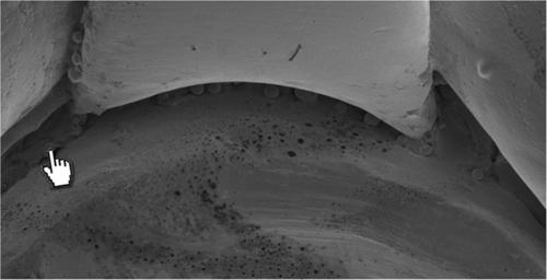
In restorative dentistry, the in situ replication of intra‐oral situations, is based on a non‐invasive and non‐destructive scanning electron microscopy (SEM) evaluation method. The technique is suitable for investigation restorative materials and dental hard‐ and soft‐tissues, and its interfaces. Surface characteristics, integrity of interfaces (margins), or fracture analysis (chipping, cracks, etc.) with reliable resolution and under high magnification (from ×50 to ×5,000). Overall the current study aims to share detailed and reproducible information about the replica technique. Specific goals are: (a) to describe detailed each step involved in producing a replica of an intra‐oral situation, (b) to validate an integrated workflow based on a rational sequence from visual examination, to macrophotography and SEM analysis using the replica technique; (c) to present three clinical cases documented using the technique. A compilation of three clinical situations/cases were analyzed here by means the replica technique showing a wide range of possibilities that can be reached and explored with the described technique. This guidance document will contribute to a more accurate use of the replica technique and help researchers and clinicians to understand and identify issues related to restorative procedures under high magnification.
中文翻译:

眼见为实?当扫描电子显微镜(SEM)符合临床牙科要求时:复制技术。
在修复性牙科中,口腔内情况的原位复制是基于无创,无损扫描电子显微镜(SEM)评估方法。该技术适用于研究修复材料以及牙齿的硬组织和软组织及其界面。具有可靠的分辨率且在高放大倍率下(从×50到×5,000)的表面特性,界面(边缘)的完整性或断裂分析(碎片,裂纹等)。总体而言,当前的研究旨在共享有关复制技术的详细且可复制的信息。具体目标是:(a)详细描述制作口腔内情况副本的每个步骤,(b)基于从外观检查到使用副本技术进行宏观摄影和SEM分析的合理顺序,验证集成的工作流程; (c)展示使用该技术记录的三个临床病例。在这里,通过复制技术分析了三种临床情况/病例的汇编,该复制技术显示了使用所述技术可以达到和探索的广泛可能性。该指导文件将有助于更准确地使用复制技术,并帮助研究人员和临床医生在高放大倍数下理解和识别与修复程序有关的问题。
更新日期:2020-07-08
中文翻译:
眼见为实?当扫描电子显微镜(SEM)符合临床牙科要求时:复制技术。
在修复性牙科中,口腔内情况的原位复制是基于无创,无损扫描电子显微镜(SEM)评估方法。该技术适用于研究修复材料以及牙齿的硬组织和软组织及其界面。具有可靠的分辨率且在高放大倍率下(从×50到×5,000)的表面特性,界面(边缘)的完整性或断裂分析(碎片,裂纹等)。总体而言,当前的研究旨在共享有关复制技术的详细且可复制的信息。具体目标是:(a)详细描述制作口腔内情况副本的每个步骤,(b)基于从外观检查到使用副本技术进行宏观摄影和SEM分析的合理顺序,验证集成的工作流程; (c)展示使用该技术记录的三个临床病例。在这里,通过复制技术分析了三种临床情况/病例的汇编,该复制技术显示了使用所述技术可以达到和探索的广泛可能性。该指导文件将有助于更准确地使用复制技术,并帮助研究人员和临床医生在高放大倍数下理解和识别与修复程序有关的问题。


 京公网安备 11010802027423号