Acta Biomaterialia ( IF 9.7 ) Pub Date : 2020-07-08 , DOI: 10.1016/j.actbio.2020.07.004 Zhi Chen 1 , Xiao Liu 1 , Jingjing You 2 , Yihui Song 2 , Eva Tomaskovic-Crook 1 , Gerard Sutton 3 , Jeremy M Crook 4 , Gordon G Wallace 1
|
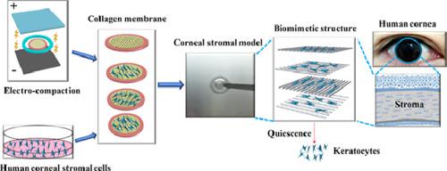
Engineering substantia propria (or stroma of cornea) that mimics the function and anatomy of natural tissue is vital for in vitro modelling and in vivo regeneration. There are, however, few examples of bioengineered biomimetic corneal stroma. Here we describe the construction of an orthogonally oriented 3D corneal stroma model (3D-CSM) using pure electro-compacted collagen (EC). EC films comprise aligned collagen fibrils and support primary human corneal stromal cells (hCSCs). Cell-laden constructs are analogous to the anatomical structure of native human cornea. The hCSCs are guided by the topographical cues provided by the aligned collagen fibrils of the EC films. Importantly, the 3D-CSM are biodegradable, highly transparent, glucose-permeable and comprise quiescent hCSCs. Gene expression analysis indicated the presence of aligned collagen fibrils is strongly coupled to downregulation of active fibroblast/myofibroblast markers α-SMA and Thy-1, with a concomitant upregulation of the dormant keratocyte marker ALDH3. The 3D-CSM represents the first example of an optimally robust biomimetic engineered corneal stroma that is constructed from pure electro-compacted collagen for cell and tissue support. The 3D-CSM is a significant advance for synthetic corneal stroma engineering, with the potential to be used for full-thickness and functional cornea replacement, as well as informing in vivo tissue regeneration.
Statement of Significance
This manuscript represents the first example of a robust, transparent, glucose permeable and pure collagen-based biomimetic 3D corneal stromal model (3D-CSM) constructed from pure electro-compacted collagen. The collagen fibrils of 3D-CSM are aligned and orthogonally arranged, mimicking native human corneal stroma. The alignment of collagen fibrils correlates with the direction of current applied for electro-compaction and influences human corneal stromal cell (hCSC) orientation. Moreover, 3D-CSM constructs support a corneal keratocyte phenotype; an essential requirement for modelling healthy corneal stroma. As-prepared 3D-CSM hold great promise as corneal stromal substitutes for research and translation, with the potential to be used for full-thickness cornea replacement.
中文翻译:
仿生角膜基质使用电致密胶原蛋白。
模仿天然组织的功能和解剖结构的工程固有质(或角膜基质)对于体外建模和体内再生至关重要。然而,几乎没有生物工程仿生角膜基质的例子。在这里,我们描述了使用纯电致密胶原(EC)构建正交定向的3D角膜基质模型(3D-CSM)。EC膜包含对齐的胶原纤维,并支持原代人角膜基质细胞(hCSC)。充满细胞的构建体类似于天然人角膜的解剖结构。hCSCs由EC膜对齐的胶原纤维提供的形貌线索指导。重要的是,3D-CSM是可生物降解的,高度透明的,葡萄糖可渗透的并且包含静态hCSC。基因表达分析表明,对齐的胶原蛋白原纤维的存在与活性成纤维细胞/成肌纤维细胞标志物α-SMA和Thy-1的下调密切相关,伴随着休眠角膜细胞标志物ALDH3的上调。3D-CSM代表了最佳耐用的仿生工程化角膜基质的第一个示例,该基质由纯电致密化胶原蛋白构建而成,可为细胞和组织提供支持。3D-CSM是合成角膜基质工程的重要进步,具有用于全厚度和功能性角膜置换以及体内组织再生的潜力。3D-CSM代表了最佳耐用的仿生工程化角膜基质的第一个示例,该基质由纯电致密化胶原蛋白构建而成,可为细胞和组织提供支持。3D-CSM是合成角膜基质工程的重要进步,具有用于全厚度和功能性角膜置换以及体内组织再生的潜力。3D-CSM代表了最佳耐用的仿生工程化角膜基质的第一个示例,该基质由纯电致密化胶原蛋白构建而成,可为细胞和组织提供支持。3D-CSM是合成角膜基质工程的重要进步,具有用于全厚度和功能性角膜置换以及体内组织再生的潜力。
重要声明
该手稿代表了由纯电致密化胶原蛋白构成的坚固,透明,葡萄糖可渗透且基于胶原蛋白的仿生3D角膜基质模型(3D-CSM)的第一个示例。3D-CSM的胶原蛋白原纤维排列并正交排列,模仿天然人类角膜基质。胶原纤维的排列与施加于电致密化的电流的方向相关,并影响人角膜基质细胞(hCSC)的方向。此外,3D-CSM构建体支持角膜角化细胞表型;建立健康角膜基质的基本要求。制备后的3D-CSM有望作为角膜基质替代物进行研究和翻译,并有望用于全厚度角膜替代。


 京公网安备 11010802027423号