当前位置:
X-MOL 学术
›
Appl. Surf. Sci.
›
论文详情
Our official English website, www.x-mol.net, welcomes your feedback! (Note: you will need to create a separate account there.)
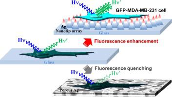
Amplified fluorescence imaging using photonic Ag nanotip array: a comparative study on surface morphology effects
Applied Surface Science ( IF 6.7 ) Pub Date : 2020-11-01 , DOI: 10.1016/j.apsusc.2020.147139 Joo-Yun Jung , Won-Geun Yang , Sin-hyoung Hong , Gun-Hwa Kim , Kiju Hwang , Weon-Sik Chae
Applied Surface Science ( IF 6.7 ) Pub Date : 2020-11-01 , DOI: 10.1016/j.apsusc.2020.147139 Joo-Yun Jung , Won-Geun Yang , Sin-hyoung Hong , Gun-Hwa Kim , Kiju Hwang , Weon-Sik Chae
|
Abstract We studied amplified fluorescence and applied to fluorescent cell imaging using plasmonic Ag substrates with the different surface nano-morphologies of tip and porous shapes. For a comparative study, we deliberately fabricated wafer-scale Ag nanotip array and nanoporous Ag substrates by a nanoimprint lithography and a chemical reduction route, respectively. Time- and space-resolved spectroscopy precisely evaluated metal-induced fluorescence characteristics by analyzing fluorescence intensity and lifetime modulations. For the standard molecular probe, the photonic Ag nanotip array showed slightly enhanced fluorescence compared to the cases on the irregular nanoporous Ag substrate and a bare glass. On the contrary, from the green fluorescent protein (GFP) expressed cell imaging study, fluorescence was remarkably amplified on the photonic nanotip array compared to that simply placed on a bare glass, whereas the GFP expressed cells suffered fluorescence quenching on the nanoporous Ag. The observed notable amplification of cell fluorescence on the photonic nanotip array is ascribed to the multiple constructive effects: the nanotip array for improved cell adherence, accumulated optical fields surrounding Ag nanotips, and photonic band-gap effect from the regularly arrayed nanotips.
中文翻译:

使用光子银纳米尖端阵列放大荧光成像:表面形态效应的比较研究
摘要 我们研究了放大的荧光,并将其应用于使用具有尖端和多孔形状的不同表面纳米形态的等离子体 Ag 基底的荧光细胞成像。为了进行比较研究,我们特意分别通过纳米压印光刻和化学还原路线制造了晶圆级 Ag 纳米尖端阵列和纳米多孔 Ag 基板。时间和空间分辨光谱通过分析荧光强度和寿命调制来精确评估金属诱导的荧光特性。对于标准分子探针,与不规则纳米多孔银基底和裸玻璃上的情况相比,光子银纳米尖端阵列显示出略微增强的荧光。相反,从绿色荧光蛋白(GFP)表达的细胞成像研究,与简单地放置在裸玻璃上相比,光子纳米尖端阵列上的荧光显着增强,而 GFP 表达的细胞在纳米多孔 Ag 上遭受荧光猝灭。在光子纳米尖端阵列上观察到的细胞荧光显着放大归因于多种建设性效应:用于改善细胞粘附的纳米尖端阵列、围绕 Ag 纳米尖端的累积光场以及来自规则排列的纳米尖端的光子带隙效应。
更新日期:2020-11-01
中文翻译:
使用光子银纳米尖端阵列放大荧光成像:表面形态效应的比较研究
摘要 我们研究了放大的荧光,并将其应用于使用具有尖端和多孔形状的不同表面纳米形态的等离子体 Ag 基底的荧光细胞成像。为了进行比较研究,我们特意分别通过纳米压印光刻和化学还原路线制造了晶圆级 Ag 纳米尖端阵列和纳米多孔 Ag 基板。时间和空间分辨光谱通过分析荧光强度和寿命调制来精确评估金属诱导的荧光特性。对于标准分子探针,与不规则纳米多孔银基底和裸玻璃上的情况相比,光子银纳米尖端阵列显示出略微增强的荧光。相反,从绿色荧光蛋白(GFP)表达的细胞成像研究,与简单地放置在裸玻璃上相比,光子纳米尖端阵列上的荧光显着增强,而 GFP 表达的细胞在纳米多孔 Ag 上遭受荧光猝灭。在光子纳米尖端阵列上观察到的细胞荧光显着放大归因于多种建设性效应:用于改善细胞粘附的纳米尖端阵列、围绕 Ag 纳米尖端的累积光场以及来自规则排列的纳米尖端的光子带隙效应。


 京公网安备 11010802027423号