当前位置:
X-MOL 学术
›
J. Neurosci. Res.
›
论文详情
Our official English website, www.x-mol.net, welcomes your
feedback! (Note: you will need to create a separate account there.)
Chronic intermittent ethanol and lipopolysaccharide exposure differentially alter Iba1-derived microglia morphology in the prelimbic cortex and nucleus accumbens core of male Long-Evans rats
Journal of Neuroscience Research ( IF 2.9 ) Pub Date : 2020-07-03 , DOI: 10.1002/jnr.24683 Benjamin M Siemsen 1, 2 , Justine D Landin 2 , Jon A McFaddin 1 , Kaylee N Hooker 1 , Lawrence J Chandler 2 , Michael D Scofield 1, 2
Journal of Neuroscience Research ( IF 2.9 ) Pub Date : 2020-07-03 , DOI: 10.1002/jnr.24683 Benjamin M Siemsen 1, 2 , Justine D Landin 2 , Jon A McFaddin 1 , Kaylee N Hooker 1 , Lawrence J Chandler 2 , Michael D Scofield 1, 2
Affiliation
|
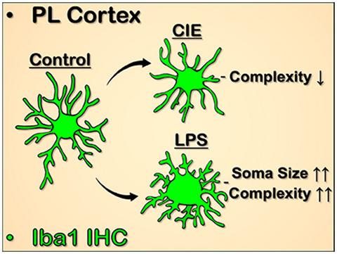
Accumulating evidence has linked pathological changes associated with chronic alcohol exposure to neuroimmune signaling mediated by microglia. Prior characterization of the microglial structure–function relationship demonstrates that alterations in activity states occur concomitantly with reorganization of cellular architecture. Accordingly, gaining a better understanding of microglial morphological changes associated with ethanol exposure will provide valuable insight into how neuroimmune signaling may contribute to ethanol-induced reshaping of neuronal function. Here we have used Iba1-staining combined with high-resolution confocal imaging and 3D reconstruction to examine microglial structure in the prelimbic (PL) cortex and nucleus accumbens (NAc) in male Long-Evans rats. Rats were either sacrificed at peak withdrawal following 15 days of exposure to chronic intermittent ethanol (CIE) or 24 hr after two consecutive injections of the immune activator lipopolysaccharide (LPS), each separated by 24 hr. LPS exposure resulted in dramatic structural reorganization of microglia in the PL cortex, including increased soma volume, overall cellular volume, and branching complexity. In comparison, CIE exposure was associated with a subtle increase in somatic volume and differential effects on microglia processes, which were largely absent in the NAc. These data reveal that microglial activation following a neuroimmune challenge with LPS or exposure to chronic alcohol exhibits distinct morphometric profiles and brain region-dependent specificity.
中文翻译:

慢性间歇性乙醇和脂多糖暴露差异改变雄性 Long-Evans 大鼠前边缘皮质和伏核核心中 Iba1 衍生的小胶质细胞形态
越来越多的证据表明,与慢性酒精暴露相关的病理变化与小胶质细胞介导的神经免疫信号有关。小胶质细胞结构-功能关系的先前表征表明,活动状态的改变与细胞结构的重组同时发生。因此,更好地了解与乙醇暴露相关的小胶质细胞形态变化将为了解神经免疫信号如何促进乙醇诱导的神经元功能重塑提供有价值的见解。在这里,我们使用 Iba1 染色结合高分辨率共聚焦成像和 3D 重建来检查雄性 Long-Evans 大鼠的前边缘 (PL) 皮层和伏隔核 (NAc) 中的小胶质细胞结构。大鼠在暴露于慢性间歇性乙醇(CIE)15天后或在连续两次注射免疫激活剂脂多糖(LPS)后24小时(每次间隔24小时)后24小时处死。LPS 暴露导致 PL 皮质中小胶质细胞发生显着的结构重组,包括体细胞体积、总体细胞体积和分支复杂性的增加。相比之下,CIE 暴露与体细胞体积的微妙增加以及对小胶质细胞过程的差异影响有关,而这在 NAc 中基本上不存在。这些数据表明,在受到 LPS 神经免疫攻击或暴露于慢性酒精后,小胶质细胞的激活表现出独特的形态特征和大脑区域依赖性特异性。
更新日期:2020-07-03
中文翻译:
慢性间歇性乙醇和脂多糖暴露差异改变雄性 Long-Evans 大鼠前边缘皮质和伏核核心中 Iba1 衍生的小胶质细胞形态
越来越多的证据表明,与慢性酒精暴露相关的病理变化与小胶质细胞介导的神经免疫信号有关。小胶质细胞结构-功能关系的先前表征表明,活动状态的改变与细胞结构的重组同时发生。因此,更好地了解与乙醇暴露相关的小胶质细胞形态变化将为了解神经免疫信号如何促进乙醇诱导的神经元功能重塑提供有价值的见解。在这里,我们使用 Iba1 染色结合高分辨率共聚焦成像和 3D 重建来检查雄性 Long-Evans 大鼠的前边缘 (PL) 皮层和伏隔核 (NAc) 中的小胶质细胞结构。大鼠在暴露于慢性间歇性乙醇(CIE)15天后或在连续两次注射免疫激活剂脂多糖(LPS)后24小时(每次间隔24小时)后24小时处死。LPS 暴露导致 PL 皮质中小胶质细胞发生显着的结构重组,包括体细胞体积、总体细胞体积和分支复杂性的增加。相比之下,CIE 暴露与体细胞体积的微妙增加以及对小胶质细胞过程的差异影响有关,而这在 NAc 中基本上不存在。这些数据表明,在受到 LPS 神经免疫攻击或暴露于慢性酒精后,小胶质细胞的激活表现出独特的形态特征和大脑区域依赖性特异性。


 京公网安备 11010802027423号