Journal of the Mechanical Behavior of Biomedical Materials ( IF 3.3 ) Pub Date : 2020-07-03 , DOI: 10.1016/j.jmbbm.2020.103935 Kazuaki Nagayama 1 , Shigeaki Ohata 1 , Shota Obata 1 , Akiko Sato 1
|
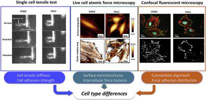
Many experimental techniques have been reported to provide knowledge of the mechanical behavior of cells from biomechanical viewpoints, however, it is unclear how the intercellular structural differences influence macroscopic and microscopic mechanical properties of cells. The aim of our study is to clarify the comprehensive mechanical properties and cell-substrate adhesion strength of cells, and the correlation with intracellular structure in different cell types. We developed an originally designed micro tensile tester, and performed a single cell tensile test to estimate whole cell tensile stiffness and adhesion strength of normal vascular smooth muscle cells (VSMCs) and cervical cancer HeLa cells: one half side of the specimen cell was lifted up by a glass microneedle, then stretched until the cell detached from the substrate, while force was simultaneously measured. The tensile stiffness and adhesion strength were 49 ± 10 nN/% and 870 ± 430 nN, respectively, in VSMCs (mean ± SD, n = 8), and 19 ± 17 nN/% and 320 ± 160 nN, respectively, in HeLa cells (n = 9). The difference was more definite in the surface elastic modulus map obtained by atomic force microscopy, indicating that the internal tension of the actin cytoskeleton was significantly higher in VSMCs than in HeLa cells. Structural analysis with confocal microscopy revealed that VSMCs had a significant alignment of F-actin cytoskeleton with mature focal adhesion, contrary to the randomly oriented F-actin with smaller focal adhesion of HeLa cells, indicating that structural arrangement of the actin cytoskeleton and their mechanical tension generated the differences in cell mechanical properties and adhesion forces. The finding strongly suggests that the mechanical and structural differences in each cell type are deeply involved with their physiological functions.
中文翻译:
使用单细胞拉伸试验和原子力显微镜对细胞的机械性能和粘附力进行宏观和微观分析:细胞类型的显着差异。
从生物力学的观点来看,已经报道了许多实验技术来提供细胞的机械行为的知识,但是,尚不清楚细胞间的结构差异如何影响细胞的宏观和微观机械性能。我们的研究目的是阐明细胞的综合机械性能和细胞-基底粘附强度,以及与不同细胞类型中细胞内结构的相关性。我们开发了最初设计的微拉伸测试仪,并进行了单细胞拉伸测试,以评估正常血管平滑肌细胞(VSMC)和宫颈癌HeLa细胞的全细胞拉伸刚度和粘附强度:将标本细胞的一半举起用玻璃微针拉伸,然后拉伸直到细胞从底物上脱落,同时测量力。在VSMC中,其拉伸刚度和粘附强度分别为49±10 nN /%和870±430 nN(均值±SD,n = 8),在HeLa中分别为19±17 nN /%和320±160 nN个单元(n = 9)。通过原子力显微镜获得的表面弹性模量图上的差异更加明确,表明VSMC中肌动蛋白细胞骨架的内部张力明显高于HeLa细胞。共聚焦显微镜的结构分析显示,VSMC具有成熟的粘着斑的F-肌动蛋白细胞骨架显着排列,与HeLa细胞的粘着力较小的随机取向的F-肌动蛋白相反,表明肌动蛋白的细胞骨架的结构排列及其机械张力产生了电池机械性能和粘附力的差异。


 京公网安备 11010802027423号