当前位置:
X-MOL 学术
›
ACS Photonics
›
论文详情
Our official English website, www.x-mol.net, welcomes your
feedback! (Note: you will need to create a separate account there.)
Nonlinear Scattering of Near-Infrared Light for Imaging Plasmonic Nanoparticles in Deep Tissue
ACS Photonics ( IF 6.5 ) Pub Date : 2020-07-01 , DOI: 10.1021/acsphotonics.0c00607 Kentaro Nishida, Gitanjal Deka, Nicholas Isaac Smith, Shi-Wei Chu, Katsumasa Fujita
ACS Photonics ( IF 6.5 ) Pub Date : 2020-07-01 , DOI: 10.1021/acsphotonics.0c00607 Kentaro Nishida, Gitanjal Deka, Nicholas Isaac Smith, Shi-Wei Chu, Katsumasa Fujita

|
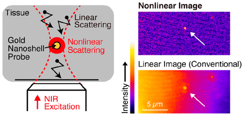
Nonlinear optical microscopy can obtain three-dimensionally resolved images within a specimen by exploiting the nonlinear light–matter interaction between the excitation light and sample. However, the image contrast significantly degrades with increasing observation depth because the emitted signal attenuates during light propagation through layers of the tissue before being detected. To obtain high contrast images from deep tissue, we developed saturated excitation (SAX) microscopy using the nonlinearity of near-infrared (NIR) plasmonic scattering from gold nanoshells and gold nanorods. SAX microscopy selectively detects the nonlinear component from the scattering signal generated by nanoparticle probes located at the center of the focal spot. By using this technique, background signals generated at out-of-focal positions are effectively removed. In addition, emitted signals in the NIR from nanoparticle probes efficiently transmit through biological tissue and are ideally suited to image deep parts of the tissue. We experimentally confirmed that scattering intensities from a single gold nanoshell and gold nanorod exhibit nonlinear relations with the excitation intensity of CW laser light at 780 and 1064 nm, respectively. We also demonstrated improvements of image contrast and spatial resolution at the depth of 400 μm in a phantom of muscle tissue by selectively detecting the nonlinear scattering signal component from gold nanoshells.
中文翻译:

近红外光的非线性散射对深部组织中的等离子体纳米粒子成像
非线性光学显微镜可以利用激发光和样品之间的非线性光-质相互作用来获得样品内的三维分辨图像。然而,图像对比度随着观察深度的增加而显着降低,这是因为所发射的信号在被检测之前穿过组织层传播的光期间会衰减。为了从深部组织获得高对比度的图像,我们利用金纳米壳和金纳米棒中近红外(NIR)等离子体散射的非线性特性开发了饱和激发(SAX)显微镜。SAX显微镜从位于焦点中心的纳米粒子探针产生的散射信号中选择性检测非线性成分。通过使用此技术,可以有效地消除在焦外位置生成的背景信号。此外,NIR中来自纳米粒子探针的发射信号有效地传输通过生物组织,并且非常适合对组织的深部成像。我们实验证实了单个金纳米壳和金纳米棒的散射强度分别与连续波激光在780 nm和1064 nm处的激发强度呈现非线性关系。我们还展示了通过选择性地检测来自金纳米壳的非线性散射信号分量,在肌肉组织模型中深度为400μm时图像对比度和空间分辨率的改善。我们实验证实了单个金纳米壳和金纳米棒的散射强度分别与连续波激光在780 nm和1064 nm处的激发强度呈现非线性关系。我们还展示了通过选择性地检测来自金纳米壳的非线性散射信号分量,在肌肉组织模型中深度为400μm时图像对比度和空间分辨率的改善。我们实验证实了单个金纳米壳和金纳米棒的散射强度分别与连续波激光在780 nm和1064 nm处的激发强度呈现非线性关系。我们还演示了通过选择性地检测来自金纳米壳的非线性散射信号分量,在肌肉组织模型中深度为400μm时图像对比度和空间分辨率的改善。
更新日期:2020-08-19
中文翻译:

近红外光的非线性散射对深部组织中的等离子体纳米粒子成像
非线性光学显微镜可以利用激发光和样品之间的非线性光-质相互作用来获得样品内的三维分辨图像。然而,图像对比度随着观察深度的增加而显着降低,这是因为所发射的信号在被检测之前穿过组织层传播的光期间会衰减。为了从深部组织获得高对比度的图像,我们利用金纳米壳和金纳米棒中近红外(NIR)等离子体散射的非线性特性开发了饱和激发(SAX)显微镜。SAX显微镜从位于焦点中心的纳米粒子探针产生的散射信号中选择性检测非线性成分。通过使用此技术,可以有效地消除在焦外位置生成的背景信号。此外,NIR中来自纳米粒子探针的发射信号有效地传输通过生物组织,并且非常适合对组织的深部成像。我们实验证实了单个金纳米壳和金纳米棒的散射强度分别与连续波激光在780 nm和1064 nm处的激发强度呈现非线性关系。我们还展示了通过选择性地检测来自金纳米壳的非线性散射信号分量,在肌肉组织模型中深度为400μm时图像对比度和空间分辨率的改善。我们实验证实了单个金纳米壳和金纳米棒的散射强度分别与连续波激光在780 nm和1064 nm处的激发强度呈现非线性关系。我们还展示了通过选择性地检测来自金纳米壳的非线性散射信号分量,在肌肉组织模型中深度为400μm时图像对比度和空间分辨率的改善。我们实验证实了单个金纳米壳和金纳米棒的散射强度分别与连续波激光在780 nm和1064 nm处的激发强度呈现非线性关系。我们还演示了通过选择性地检测来自金纳米壳的非线性散射信号分量,在肌肉组织模型中深度为400μm时图像对比度和空间分辨率的改善。


 京公网安备 11010802027423号