Spectrochimica Acta Part A: Molecular and Biomolecular Spectroscopy ( IF 4.3 ) Pub Date : 2020-07-02 , DOI: 10.1016/j.saa.2020.118666 P Sakthivel 1 , K Kavi Rasu 1 , G K D Prasanna Venkatesan 2 , Amelec Viloria 3
|
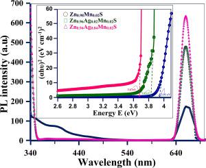
The current study deals with the structural, morphological, elemental, optical and photoluminescence behaviors of Ag+, Mn2+ dual doped ZnS quantum dots (QDs). The X-ray diffraction (XRD) and Transmission Electron Microscope (TEM) studies confirmed the cubic structure and size of the crystallites (~2 nm). The Scanning Electron Microscope (SEM) photographs portrayed the surface and morphological structure of prepared samples. Energy dispersive X-ray (EDX) and Fourier Transform Infrared Spectra (FTIR) ensured the presence of Zn, Ag, Mn and, S in the samples as per the anticipated stoichiometry ratio. The UV–visible spectra showed a red shift in optical absorption and band gap gets narrowed due to the incorporation of Ag+ ions. The size effect has overcome the quantum confinement effect in this case. Through photoluminescence (PL) studies, a weak UV emission and strong red wavelength emissions were received and discussed on the basis of sulfur vacancies. This red emission was dealt in terms of d-electrons transition between host and dopant ions.
中文翻译:
Ag +和Mn2 +离子对ZnS量子点的结构,光学和光致发光特性的影响。
当前的研究涉及Ag +,Mn 2+双掺杂ZnS量子点(QDs)的结构,形态,元素,光学和光致发光行为。X射线衍射(XRD)和透射电子显微镜(TEM)研究证实了微晶的立方结构和尺寸(约2 nm)。扫描电子显微镜(SEM)的照片描绘了所制备样品的表面和形态结构。能量色散X射线(EDX)和傅里叶变换红外光谱(FTIR)可确保按照预期的化学计量比在样品中存在Zn,Ag,Mn和S。紫外可见光谱显示,由于Ag +的引入,光吸收发生红移,并且带隙变窄离子。在这种情况下,尺寸效应克服了量子限制效应。通过光致发光(PL)研究,在硫空位的基础上收到并讨论了弱UV发射和强红色波长发射。根据主体离子和掺杂离子之间的d电子跃迁处理了这种红色发射。


 京公网安备 11010802027423号