当前位置:
X-MOL 学术
›
Biomater. Sci.
›
论文详情
Our official English website, www.x-mol.net, welcomes your
feedback! (Note: you will need to create a separate account there.)
An RGD modified water-soluble fluorophore probe for in vivo NIR-II imaging of thrombosis.
Biomaterials Science ( IF 5.8 ) Pub Date : 2020-07-01 , DOI: 10.1039/d0bm00729c Yanping Wu 1 , Chao Wang , Jiaqi Guo , Abdlay Carvalho , Yuyu Yao , Pengfei Sun , Quli Fan
Biomaterials Science ( IF 5.8 ) Pub Date : 2020-07-01 , DOI: 10.1039/d0bm00729c Yanping Wu 1 , Chao Wang , Jiaqi Guo , Abdlay Carvalho , Yuyu Yao , Pengfei Sun , Quli Fan
Affiliation
|
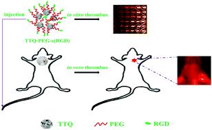
Venous thrombosis leads to severe symptoms and death through pulmonary embolism. There is a great need for high sensitivity imaging methods to identify acute patients who would benefit from thrombolysis. We designed a novel, organic near-infrared second-window (NIR-II) probe, which targets the glycoprotein IIb/IIIa receptor (GPIIb/IIIa) on activated platelets. The probe's structure was characterized by MALDI-TOF-MS, TEM, UV-visible absorption and NIR-II fluorescent spectroscopy. The probe's specificity for activated platelets was investigated in vitro and in vivo. Thrombosis in mice was induced by administration of FeCl3 in the external jugular vein and imaged by using a NIR-II imager. The donor–acceptor–donor fluorescent core TTQ was prepared from donor and acceptor units. TTQ-PEG-NH2 was synthesized by sequential modification of PEGylated TTQ, followed by c(RGD) condensation. Signal strength was continuously monitored for 24 h following TTQ-PEG-c(RGD) or non-specific fluorescent dye injection. The contralateral external jugular vein, sham surgery and a competitive inhibition experiment served as controls. TTQ-PEG-c(RGD) presented high NIR-II intensity, good stability and excellent affinity for activated platelets. The NIR-II fluorescence signal of TTQ-PEG-c(RGD) injected mice significantly increased at the thrombus site and peaked at 4 h, whereas there was no significant change in the control mice, and the competitive inhibition of the RGD antagonist suppressed the enhancement of the NIR-II fluorescence signal. Comparison between fresh and old thrombi confirmed that TTQ-PEG-c(RGD) could be used to distinguish a fresh thrombus from an old thrombus. TTQ-PEG-c(RGD) can specifically target thrombosis in vitro and in vivo, providing a potential tool for noninvasive diagnosis of early thrombi.
中文翻译:

RGD修饰的水溶性荧光团探针,用于体内血栓形成的NIR-II成像。
静脉血栓形成通过肺栓塞导致严重的症状和死亡。迫切需要高灵敏度的影像学方法来识别可从溶栓治疗中受益的急性患者。我们设计了一种新颖的,有机的近红外第二窗口(NIR-II)探针,其靶向活化血小板上的糖蛋白IIb / IIIa受体(GPIIb / IIIa)。探针的结构通过MALDI-TOF-MS,TEM,紫外可见吸收和NIR-II荧光光谱进行了表征。在体外和体内研究了该探针对活化血小板的特异性。给予FeCl 3可诱发小鼠血栓形成在颈外静脉中成像,并使用NIR-II成像仪成像。供体-受体-供体荧光核心TTQ由供体和受体单元制备。TTQ-PEG-NH 2通过依次修饰聚乙二醇化的TTQ,然后进行c(RGD)缩合来合成α-己内酰胺。在注入TTQ-PEG-c(RGD)或非特异性荧光染料后,连续24 h监测信号强度。将对侧颈外静脉,假手术和竞争性抑制实验作为对照。TTQ-PEG-c(RGD)具有高NIR-II强度,良好的稳定性和对活化血小板的优异亲和力。注射TTQ-PEG-c(RGD)的小鼠的NIR-II荧光信号在血栓部位显着增加并在4 h达到峰值,而对照小鼠则没有明显变化,RGD拮抗剂的竞争性抑制作用抑制了该作用。 NIR-II荧光信号的增强。新旧血栓之间的比较证实,TTQ-PEG-c(RGD)可用于区分新鲜血栓和旧血栓。TTQ-PEG-c(RGD)可以特异性靶向血栓形成体外和体内,为早期血栓的无创诊断提供了潜在的工具。
更新日期:2020-08-11
中文翻译:
RGD修饰的水溶性荧光团探针,用于体内血栓形成的NIR-II成像。
静脉血栓形成通过肺栓塞导致严重的症状和死亡。迫切需要高灵敏度的影像学方法来识别可从溶栓治疗中受益的急性患者。我们设计了一种新颖的,有机的近红外第二窗口(NIR-II)探针,其靶向活化血小板上的糖蛋白IIb / IIIa受体(GPIIb / IIIa)。探针的结构通过MALDI-TOF-MS,TEM,紫外可见吸收和NIR-II荧光光谱进行了表征。在体外和体内研究了该探针对活化血小板的特异性。给予FeCl 3可诱发小鼠血栓形成在颈外静脉中成像,并使用NIR-II成像仪成像。供体-受体-供体荧光核心TTQ由供体和受体单元制备。TTQ-PEG-NH 2通过依次修饰聚乙二醇化的TTQ,然后进行c(RGD)缩合来合成α-己内酰胺。在注入TTQ-PEG-c(RGD)或非特异性荧光染料后,连续24 h监测信号强度。将对侧颈外静脉,假手术和竞争性抑制实验作为对照。TTQ-PEG-c(RGD)具有高NIR-II强度,良好的稳定性和对活化血小板的优异亲和力。注射TTQ-PEG-c(RGD)的小鼠的NIR-II荧光信号在血栓部位显着增加并在4 h达到峰值,而对照小鼠则没有明显变化,RGD拮抗剂的竞争性抑制作用抑制了该作用。 NIR-II荧光信号的增强。新旧血栓之间的比较证实,TTQ-PEG-c(RGD)可用于区分新鲜血栓和旧血栓。TTQ-PEG-c(RGD)可以特异性靶向血栓形成体外和体内,为早期血栓的无创诊断提供了潜在的工具。


 京公网安备 11010802027423号