当前位置:
X-MOL 学术
›
J. Raman Spectrosc.
›
论文详情
Our official English website, www.x-mol.net, welcomes your
feedback! (Note: you will need to create a separate account there.)
Feasibility of integrated high‐wavenumber Raman imaging and fingerprint Raman spectroscopy for fast margin assessment in breast cancer surgery
Journal of Raman Spectroscopy ( IF 2.4 ) Pub Date : 2020-06-30 , DOI: 10.1002/jrs.5937 Zhiyu Liao 1 , Maria Giovanna Lizio 1 , Christopher Corden 1 , Hazem Khout 2 , Emad Rakha 3 , Ioan Notingher 1
Journal of Raman Spectroscopy ( IF 2.4 ) Pub Date : 2020-06-30 , DOI: 10.1002/jrs.5937 Zhiyu Liao 1 , Maria Giovanna Lizio 1 , Christopher Corden 1 , Hazem Khout 2 , Emad Rakha 3 , Ioan Notingher 1
Affiliation
|
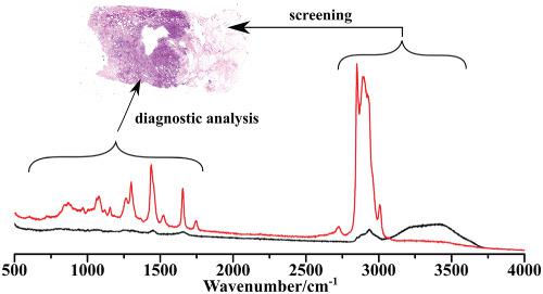
Intraoperative assessment of surgical margins remains one of the main challenges in cancer surgery. Raman spectroscopy can detect cancer cells with high accuracy, but it is time‐consuming. In this paper, we investigated a selective‐sampling Raman spectroscopy approach, based on high wavenumber (HW) Raman imaging (spectral range 2,500–3,500 cm−1) and fingerprint Raman spectroscopy (spectral range 600–1,800 cm−1), to reduce the overall tissue analysis time while maintaining high diagnostic accuracy. HW Raman mapping was used as a first step to identify the adipose tissue regions based on the C–H stretching bands at 2,700–2,950 cm−1. As residual tumors are typically found in nonadipose tissue, an algorithm was developed to allocate sampling points for fingerprint Raman spectroscopy at locations corresponding to low intensity in the HW‐Raman maps. Preliminary results show that HW‐Raman imaging based on a 671 nm laser is effective and fast for mapping of adipose tissue in breast resections, with typical imaging times of 2 min for tissue areas as large as 2 × 2 cm2 areas. Albeit the remaining high fluorescence background in the fingerprint region prevents the use of single 671‐nm laser, the HW Raman imaging can be still exploited in combination with 785‐nm excitation Raman spectroscopy for identifying residual tumor. Although this study demonstrates the feasibility of this approach, further improvements, such as using single element detectors for HW Raman imaging, are required to increase the analysis speed further towards intraoperative use in the routine clinical setting.
中文翻译:

集成高波数拉曼成像和指纹拉曼光谱在乳腺癌手术中快速评估边缘的可行性
术中对手术切缘的评估仍然是癌症手术中的主要挑战之一。拉曼光谱法可以高精度检测癌细胞,但是很费时。在本文中,我们研究了基于高波数(HW)拉曼成像(光谱范围2,500–3,500 cm -1)和指纹拉曼光谱(光谱范围600–1,800 cm -1)的选择性采样拉曼光谱方法,以减少总体组织分析时间,同时保持较高的诊断准确性。HW拉曼作图被用作基于2700–2,950 cm -1处C–H拉伸带识别脂肪组织区域的第一步。由于通常在非脂肪组织中发现残留肿瘤,因此开发了一种算法,可在与HW-Raman图中低强度相对应的位置分配用于指纹拉曼光谱的采样点。初步结果表明,基于671 nm激光的HW‐Raman成像对乳腺切除术中的脂肪组织进行制图是有效且快速的,对于2×2 cm 2的组织区域,典型成像时间为2分钟地区。尽管指纹区域中剩余的高荧光背景无法使用单个671 nm激光,但仍然可以将HW拉曼成像与785 nm激发拉曼光谱结合使用以鉴定残留肿瘤。尽管这项研究证明了这种方法的可行性,但仍需要进一步的改进,例如使用单元素检测器进行HW拉曼成像,以提高在常规临床环境中术中使用时的分析速度。
更新日期:2020-06-30
中文翻译:
集成高波数拉曼成像和指纹拉曼光谱在乳腺癌手术中快速评估边缘的可行性
术中对手术切缘的评估仍然是癌症手术中的主要挑战之一。拉曼光谱法可以高精度检测癌细胞,但是很费时。在本文中,我们研究了基于高波数(HW)拉曼成像(光谱范围2,500–3,500 cm -1)和指纹拉曼光谱(光谱范围600–1,800 cm -1)的选择性采样拉曼光谱方法,以减少总体组织分析时间,同时保持较高的诊断准确性。HW拉曼作图被用作基于2700–2,950 cm -1处C–H拉伸带识别脂肪组织区域的第一步。由于通常在非脂肪组织中发现残留肿瘤,因此开发了一种算法,可在与HW-Raman图中低强度相对应的位置分配用于指纹拉曼光谱的采样点。初步结果表明,基于671 nm激光的HW‐Raman成像对乳腺切除术中的脂肪组织进行制图是有效且快速的,对于2×2 cm 2的组织区域,典型成像时间为2分钟地区。尽管指纹区域中剩余的高荧光背景无法使用单个671 nm激光,但仍然可以将HW拉曼成像与785 nm激发拉曼光谱结合使用以鉴定残留肿瘤。尽管这项研究证明了这种方法的可行性,但仍需要进一步的改进,例如使用单元素检测器进行HW拉曼成像,以提高在常规临床环境中术中使用时的分析速度。


 京公网安备 11010802027423号