当前位置:
X-MOL 学术
›
J. Morphol.
›
论文详情
Our official English website, www.x-mol.net, welcomes your
feedback! (Note: you will need to create a separate account there.)
The brain structure of selected trypanorhynch tapeworms
Journal of Morphology ( IF 1.5 ) Pub Date : 2020-06-30 , DOI: 10.1002/jmor.21145 Natalia M Biserova 1 , Janetta V Korneva 2 , Tatiana A Polyakova 3
Journal of Morphology ( IF 1.5 ) Pub Date : 2020-06-30 , DOI: 10.1002/jmor.21145 Natalia M Biserova 1 , Janetta V Korneva 2 , Tatiana A Polyakova 3
Affiliation
|
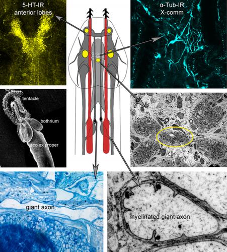
The brain architecture in four species of tapeworms from the order Trypanorhyncha has been studied. In all species, the brain consists of paired anterior and lateral lobes, and an unpaired central lobe. The anterior lobes connect by dorsal and ventral semicircular commissures; the central and lateral lobes connect by a median and an X‐shaped crisscross commissure. In the center of the brain, five well‐developed compact neuropils are present. The brain occupies a medial position in the scolex pars bothrialis. The ventral excretory vessels are situated outside the lateral lobes of the brain; the dorsal excretory vessels are located inside the brain and dorsal to the median commissure. The brain gives rize four anterior proboscis nerves and four posterior bulbar nerves with myelinated giant axons (GAs). The cell bodies of the GAs are located within the X‐commissure and in the bulbar nerves. Highly developed serotonergic neuropils are present in the anterior and lateral lobes; numerous 5‐HT neurons are found in the brain lobes including the central unpaired lobe. The X‐cross commissure consists of the α‐tub‐immunoreactive and 5‐HT‐IR neurites. Eight ultrastructural types of neurons were found in the brain of the three species investigated. In addition, different types of synapses were present in the neuropils. Glial cells ensheath the brain lobes, the neuropils, the GAs, and the bulbar nerves. Glia cell processes form complex branching patterns of thin cytoplasmic sheets sandwiched between adjacent neural processes and filling the space between neurons. Multilayer myelin‐like envelopes and a mesaxon‐like structure have been found in Trypanorhyncha nervous system. We compared the brain architecture of Trypanorhyncha with that of an early basal cestode taxon, that is, Diphyllobothriidea, and present a hypothesis about the homology of the anterior brain lobes in order Trypanorhyncha; and the lateral lobes and median commissure are homologous brain structures within Eucestoda.
中文翻译:

选定的锥虫绦虫的大脑结构
已经研究了来自锥虫目的四种绦虫的大脑结构。在所有物种中,大脑由成对的前叶和侧叶以及不成对的中央叶组成。前叶由背侧和腹侧半圆形连合处连接;中央叶和侧叶由中叶和 X 形交叉连合处连接。在大脑中央,存在五个发育良好的致密神经纤维。大脑在头节中占据中间位置。腹侧排泄血管位于大脑侧叶外;背侧排泄血管位于大脑内部和正中连合背侧。大脑产生四根前鼻神经和四根后延髓神经,带有髓鞘巨轴突 (GA)。GA 的细胞体位于 X 连合内和延髓神经内。前叶和侧叶存在高度发达的血清素能神经细胞;在包括中央未配对脑叶在内的脑叶中发现了许多 5-HT 神经元。X-cross commissure 由 α-tub-immunoreactive 和 5-HT-IR 轴突组成。在所研究的三个物种的大脑中发现了八种超微结构类型的神经元。此外,神经元中还存在不同类型的突触。神经胶质细胞包裹着脑叶、神经细胞、GA 和延髓神经。神经胶质细胞突起形成夹在相邻神经突起之间并填充神经元之间空间的薄细胞质片的复杂分支模式。在锥虫神经系统中发现了多层髓鞘样包膜和中轴样结构。我们将锥虫的脑结构与早期基底绦虫分类群(即 Diphyllobothriidea)的脑结构进行了比较,并提出了关于锥虫的前脑叶同源性的假设;侧叶和正中连合是 Eucestoda 内的同源大脑结构。
更新日期:2020-06-30
中文翻译:
选定的锥虫绦虫的大脑结构
已经研究了来自锥虫目的四种绦虫的大脑结构。在所有物种中,大脑由成对的前叶和侧叶以及不成对的中央叶组成。前叶由背侧和腹侧半圆形连合处连接;中央叶和侧叶由中叶和 X 形交叉连合处连接。在大脑中央,存在五个发育良好的致密神经纤维。大脑在头节中占据中间位置。腹侧排泄血管位于大脑侧叶外;背侧排泄血管位于大脑内部和正中连合背侧。大脑产生四根前鼻神经和四根后延髓神经,带有髓鞘巨轴突 (GA)。GA 的细胞体位于 X 连合内和延髓神经内。前叶和侧叶存在高度发达的血清素能神经细胞;在包括中央未配对脑叶在内的脑叶中发现了许多 5-HT 神经元。X-cross commissure 由 α-tub-immunoreactive 和 5-HT-IR 轴突组成。在所研究的三个物种的大脑中发现了八种超微结构类型的神经元。此外,神经元中还存在不同类型的突触。神经胶质细胞包裹着脑叶、神经细胞、GA 和延髓神经。神经胶质细胞突起形成夹在相邻神经突起之间并填充神经元之间空间的薄细胞质片的复杂分支模式。在锥虫神经系统中发现了多层髓鞘样包膜和中轴样结构。我们将锥虫的脑结构与早期基底绦虫分类群(即 Diphyllobothriidea)的脑结构进行了比较,并提出了关于锥虫的前脑叶同源性的假设;侧叶和正中连合是 Eucestoda 内的同源大脑结构。


 京公网安备 11010802027423号