当前位置:
X-MOL 学术
›
J. Biophotonics
›
论文详情
Our official English website, www.x-mol.net, welcomes your
feedback! (Note: you will need to create a separate account there.)
Raman microspectroscopic study for the detection of oral field cancerisation using brush biopsy samples.
Journal of Biophotonics ( IF 2.0 ) Pub Date : 2020-06-29 , DOI: 10.1002/jbio.202000131 Isha Behl 1, 2 , Genecy Calado 1, 2 , Anika Vishwakarma 1, 2 , Stephen Flint 3 , Sheila Galvin 3 , Claire M Healy 3 , Marina Leite Pimentel 4 , Alison Malkin 5 , Hugh J Byrne 6 , Fiona M Lyng 1, 2
Journal of Biophotonics ( IF 2.0 ) Pub Date : 2020-06-29 , DOI: 10.1002/jbio.202000131 Isha Behl 1, 2 , Genecy Calado 1, 2 , Anika Vishwakarma 1, 2 , Stephen Flint 3 , Sheila Galvin 3 , Claire M Healy 3 , Marina Leite Pimentel 4 , Alison Malkin 5 , Hugh J Byrne 6 , Fiona M Lyng 1, 2
Affiliation
|
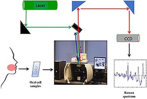
Field cancerisation (FC) is potentially an underlying cause of poor treatment outcomes of oral squamous cell carcinoma (OSCC). To explore the phenomenon using Raman microspectroscopy, brush biopsies from the buccal mucosa, tongue, gingiva and alveolus of healthy donors (n = 40) and from potentially malignant lesions (PML) of Dysplasia Clinic patients (n = 40) were examined. Contralateral normal samples (n = 38) were also collected from the patients. Raman spectra were acquired from the nucleus and cytoplasm of each cell, and subjected to partial least squares‐discriminant analysis (PLS‐DA). High discriminatory accuracy for donor and PML samples was achieved for both cytopalmic and nuclear data sets. Notably, contralateral normal (patient) samples were also accurately discriminated from donor samples and contralateral normal samples from patients with multiple lesions showed a similar spectral profile to PML samples, strongly indicating a FC effect. These findings support the potential of Raman microspectroscopy as a screening tool for PML using oral exfoliated cells.
中文翻译:

拉曼显微光谱研究,用于使用刷式活检样本检测口腔野癌。
现场癌变(FC)可能是口腔鳞状细胞癌(OSCC)治疗效果不佳的根本原因。为了使用拉曼光谱研究该现象,对健康供体的颊黏膜,舌头,齿龈和肺泡(n = 40)以及发育不良诊所患者(n = 40)的潜在恶性病变(PML)进行了活检。还从患者中收集了对侧正常样本(n = 38)。拉曼光谱是从每个细胞的细胞核和细胞质中获取的,并进行了偏最小二乘判别分析(PLS-DA)。对于细胞周期和核数据集,对供体和PML样品均实现了较高的区分精度。值得注意的是 对侧正常(患者)样本也可以与供体样本准确区分开,多个病变患者的对侧正常样本的光谱特征与PML样本相似,强烈表明有FC作用。这些发现支持了拉曼显微术作为使用口腔脱落细胞的PML筛查工具的潜力。
更新日期:2020-06-29
中文翻译:
拉曼显微光谱研究,用于使用刷式活检样本检测口腔野癌。
现场癌变(FC)可能是口腔鳞状细胞癌(OSCC)治疗效果不佳的根本原因。为了使用拉曼光谱研究该现象,对健康供体的颊黏膜,舌头,齿龈和肺泡(n = 40)以及发育不良诊所患者(n = 40)的潜在恶性病变(PML)进行了活检。还从患者中收集了对侧正常样本(n = 38)。拉曼光谱是从每个细胞的细胞核和细胞质中获取的,并进行了偏最小二乘判别分析(PLS-DA)。对于细胞周期和核数据集,对供体和PML样品均实现了较高的区分精度。值得注意的是 对侧正常(患者)样本也可以与供体样本准确区分开,多个病变患者的对侧正常样本的光谱特征与PML样本相似,强烈表明有FC作用。这些发现支持了拉曼显微术作为使用口腔脱落细胞的PML筛查工具的潜力。


 京公网安备 11010802027423号