当前位置:
X-MOL 学术
›
Adv. Healthcare Mater.
›
论文详情
Our official English website, www.x-mol.net, welcomes your
feedback! (Note: you will need to create a separate account there.)
An Oxidation-Enhanced Magnetic Resonance Imaging Probe for Visual and Specific Detection of Singlet Oxygen Generated in Photodynamic Cancer Therapy In Vivo.
Advanced Healthcare Materials ( IF 10.0 ) Pub Date : 2020-06-30 , DOI: 10.1002/adhm.202000533 Kai Deng 1 , Bo Wu 1 , Cai-Xia Wang 1 , Qian Wang 1 , Hui Yu 1 , Jia-Mi Li 1 , Kun-Heng Li 1 , Hong-Yang Zhao 1 , Shi-Wen Huang 1
Advanced Healthcare Materials ( IF 10.0 ) Pub Date : 2020-06-30 , DOI: 10.1002/adhm.202000533 Kai Deng 1 , Bo Wu 1 , Cai-Xia Wang 1 , Qian Wang 1 , Hui Yu 1 , Jia-Mi Li 1 , Kun-Heng Li 1 , Hong-Yang Zhao 1 , Shi-Wen Huang 1
Affiliation
|
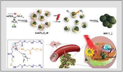
Singlet oxygen is regarded as the primary cytotoxic agent in cancer photodynamic therapy (PDT). Despite the advances in optical methods to image singlet oxygen, it remains a challenge for in vivo application due to the limited tissue penetration depth of light. Up to date, no singlet oxygen‐specific magnetic resonance imaging (MRI) probe has been reported. Herein, a T2‐weighted MRI probe is reported to visually detect singlet oxygen generated in PDT in vitro and in vivo. The MRI probe Ce6/Fe3O4‐M is constructed by co‐encapsulation of photosensitizer Ce6 and Fe3O4 nanoparticles in mPEG2000‐TK‐C16 micelles. Thioketal (TK) linker in the probe is highly sensitive to singlet oxygen, but lowly sensitive to other reactive oxygen species (ROS) existing in physiological and pathological environments. Singlet oxygen, generated with light irradiation, triggers the cleavage of TK, which leads to loss of surface polyethylene glycol, increment of the hydrophobicity, and aggregation of Fe3O4 nanoparticles. Subsequently, negatively enhanced T2‐weighted MRI signal is obtained for visual detection of singlet oxygen in the solution, cancer cells, and in vivo. This oxidation responsive MRI probe is expected to hold great promise in evaluating the ability of photosensitizers to generate singlet oxygen and in predicting the therapeutic efficacies of PDT in vivo.
中文翻译:

氧化增强磁共振成像探针,用于视觉和特异性检测体内光动力癌症治疗中产生的单线态氧。
单线态氧被认为是癌症光动力疗法(PDT)中的主要细胞毒剂。尽管光学方法用于对单线态氧进行成像,但由于光的组织穿透深度有限,对于体内应用仍然是一个挑战。迄今为止,尚无单线态氧特异性磁共振成像(MRI)探针的报道。本文中,据报道,T 2加权MRI探针可在体外和体内目视检测PDT中产生的单线态氧。MRI探针Ce6 / Fe 3 O 4‐ M是通过将光敏剂Ce6和Fe 3 O 4纳米粒子共包裹在mPEG 2000 ‐TK‐C 16中构建的胶束。探针中的Thioketal(TK)接头对单线态氧高度敏感,但对生理和病理环境中存在的其他活性氧种类(ROS)则较不敏感。光辐射产生的单线态氧触发TK的裂解,从而导致表面聚乙二醇损失,疏水性增加和Fe 3 O 4纳米粒子聚集。随后,T 2负增强获得加权的MRI信号,以可视方式检测溶液,癌细胞和体内的单线态氧。这种氧化反应性MRI探针有望在评估光敏剂产生单线态氧的能力以及预测PDT在体内的治疗效果方面具有广阔的前景。
更新日期:2020-08-19
中文翻译:
氧化增强磁共振成像探针,用于视觉和特异性检测体内光动力癌症治疗中产生的单线态氧。
单线态氧被认为是癌症光动力疗法(PDT)中的主要细胞毒剂。尽管光学方法用于对单线态氧进行成像,但由于光的组织穿透深度有限,对于体内应用仍然是一个挑战。迄今为止,尚无单线态氧特异性磁共振成像(MRI)探针的报道。本文中,据报道,T 2加权MRI探针可在体外和体内目视检测PDT中产生的单线态氧。MRI探针Ce6 / Fe 3 O 4‐ M是通过将光敏剂Ce6和Fe 3 O 4纳米粒子共包裹在mPEG 2000 ‐TK‐C 16中构建的胶束。探针中的Thioketal(TK)接头对单线态氧高度敏感,但对生理和病理环境中存在的其他活性氧种类(ROS)则较不敏感。光辐射产生的单线态氧触发TK的裂解,从而导致表面聚乙二醇损失,疏水性增加和Fe 3 O 4纳米粒子聚集。随后,T 2负增强获得加权的MRI信号,以可视方式检测溶液,癌细胞和体内的单线态氧。这种氧化反应性MRI探针有望在评估光敏剂产生单线态氧的能力以及预测PDT在体内的治疗效果方面具有广阔的前景。


 京公网安备 11010802027423号