Surface & Coatings Technology ( IF 5.3 ) Pub Date : 2020-06-30 , DOI: 10.1016/j.surfcoat.2020.126125 Chiang-Sang Chen , Jean-Heng Chang , Viritpon Srimaneepong , Jia-Yi Wen , Oi-Hong Tung , Chih-Hsiung Yang , Hui-Ching Lin , Tszu-Hsin Lee , Yong Han , Her-Hsiung Huang
|
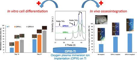
Titanium (Ti) is widely used in dental implants; however, the bioinert surface has been shown to limit osseointegration, particularly in patients with poor bone quality. This study created a thin bioactive oxide film on Ti dental implants via oxygen plasma immersion ion implantation (OPIII) to improve bone cell differentiation and osseointegration. OPIII treatment was performed using low (1016 ions/cm2) and high (4 × 10¹⁷ ions/cm2) doses of oxygen ions. The Ti surfaces were characterized in terms of topography, wettability, chemical composition, and crystal structure. The differentiation of human bone marrow mesenchymal stem cells on OPIII-treated Ti surfaces was evaluated in vitro in terms of alkaline phosphatase (ALP) activity (early stage marker) and von Kossa staining (late stage marker). The osseointegration of OPIII-treated screw-type Ti dental implants in the femur of rabbits was evaluated in vivo using scanning electron microscope/backscattered electron imaging and histological staining at 4 weeks after implantation. OPIII treatment could induce a Ti oxide film on Ti surface without altering the surface topography, roughness or wettability. Higher oxygen ions dose increased the thickness of the surface Ti oxide film as well as the proportion of rutile phase titanium dioxide (TiO2) in the Ti oxide film. The OPIII-treated Ti surfaces presented higher ALP expression level, stronger von Kossa staining signal, and higher bone-to-implant contact. Note that all of these effects were more pronounced on surfaces created using higher oxygen ions dose due to the higher proportion of rutile phase TiO2. Overall, our results indicate that OPIII treatment using higher oxygen ions dose can enhance the in vitro bone cell differentiation and in vivo osseointegration of Ti dental implants.
中文翻译:
提高了体外细胞分化和体内通过氧钛牙科植入物骨整合的等离子体浸没离子注入处理
钛(Ti)广泛用于牙科植入物;然而,已证明生物惰性表面会限制骨整合,特别是在骨质较差的患者中。这项研究通过氧等离子体浸入离子植入(OPIII)在Ti牙科植入物上创建了一层薄薄的生物活性氧化物膜,以改善骨细胞分化和骨整合。使用低剂量(10 16 离子/ cm 2)和高剂量(4×10 17 离子/ cm 2)的氧离子进行OPIII处理。Ti表面的形貌,润湿性,化学组成和晶体结构均得到了表征。体外评估OPIII处理的Ti表面上人骨髓间充质干细胞的分化就碱性磷酸酶(ALP)活性(早期标记)和冯·科萨染色(晚期标记)而言。在植入后第4周,使用扫描电子显微镜/反向散射电子成像和组织学染色在体内评估了用OPIII处理的螺钉型Ti牙科植入物在兔股骨中的骨整合。OPIII处理可在不改变表面形貌,粗糙度或润湿性的情况下在Ti表面诱导Ti氧化膜。较高的氧离子剂量会增加表面Ti氧化膜的厚度以及金红石相二氧化钛(TiO 2)在氧化钛膜中。OPIII处理的Ti表面表现出更高的ALP表达水平,更强的von Kossa染色信号以及更高的骨骼与植入物接触。请注意,由于金红石相TiO 2的比例较高,因此在使用较高的氧离子剂量创建的表面上,所有这些效果均更为明显。总体而言,我们的结果表明,使用更高的氧离子剂量进行OPIII治疗可以增强Ti牙植入物的体外骨细胞分化和体内骨整合。


 京公网安备 11010802027423号