Journal of Neuroscience Methods ( IF 2.7 ) Pub Date : 2020-06-30 , DOI: 10.1016/j.jneumeth.2020.108838 Kyung Sik Yi 1 , Chi-Hoon Choi 1 , Cheolkyu Jung 2 , Youngjeon Lee 3 , Chang-Yeop Jeon 3 , Hyun-Gu Yeo 3 , Yujin Ahn 3 , Jinwoo Hwang 4 , Hong Jun Lee 5 , Janggeun Cho 6 , Bitnarae Kwak 6 , Kyung A Kwak 6 , Sang-Rae Lee 3 , Sang-Hoon Cha 7
|
Background
The study aimed to establish a staining method that could delineate the macroscopic lesion boundary of a hyperacute infarction depicted by diffusion-weighted MRI (DWI) and to validate the infarction boundary by comparing different staining methods.
New Method
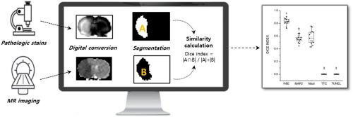
Thirteen rats with 1 -h middle cerebral artery (MCA) infarction were included. Five different staining methods (Hematoxylin and eosin (H&E), Nissl, 2,3,5-triphenyltetrazolium hydrochloride (TTC), microtubule associated protein 2 (MAP2), and terminal deoxynucleotidyl transferase dUTP nick end labeling (TUNEL) stains) were used to identify whether the hyperacute infarction could be histopathologically identified. Dice indices were compared to evaluate similarities in the lesion area ascertained by DWI and the staining methods. Through macroscopic lesion delineation, each region was subdivided into abnormal regions in all three stains (ROIA), abnormal in two stains (ROIB), and abnormal in only one (ROIC). Microscopic cellular changes were evaluated and graded according to each region.
Results
Mean Dice indices of the H&E stain were significantly higher than those of the Nissl- and MAP2-stained specimens (0.83 ± 0.052, 0.58 ± 0.107, and 0.56 ± 0.059, respectively; p = 0.000). Grading scores for ROIs in the DWI abnormal lesions varied by region: ROIA exhibited the most severe damage [median (IQR), 3 (1)], followed respectively by ROIB [median (IQR), 2 (0)] and ROIC [median (IQR), 1 (0)]
Comparison with existing methods
H&E stain best reflects 1 h hyperacute DWI abnormal lesions.
Conclusions
H&E stain allowed for the macroscopic delineation of the 1 h DWI-abnormal lesions, while MAP2 and Nissl stains could only partially depict lesions.
中文翻译:
哪种病理染色方法可以可视化通过扩散MRI识别的超急性梗死灶?:一项对比实验研究。
背景
这项研究旨在建立一种染色方法,以描绘弥散加权MRI(DWI)描绘的超急性梗死的宏观病变边界,并通过比较不同的染色方法来验证梗死边界。
新方法
包括13只1小时脑中动脉(MCA)梗塞的大鼠。五种不同的染色方法(苏木精和曙红(H&E),尼氏,2,3,5-三苯基四唑盐酸盐(TTC),微管相关蛋白2(MAP2)和末端脱氧核苷酸转移酶dUTP缺口末端标记(TUNEL))确定是否可以在组织病理学上鉴定超急性梗死。比较骰子指数以评估通过DWI和染色方法确定的病变区域的相似性。通过宏观病灶划分,每个区域被细分为异常区域在所有三个污渍(ROI甲)在两个污渍(ROI异常乙),和异常仅在一个(ROI Ç)。根据每个区域评估微观细胞变化并分级。
结果
H&E染色的平均Dice指数显着高于Nissl和MAP2染色的样本(分别为0.83±0.052、0.58±0.107和0.56±0.059; p = 0.000)。DWI异常病变中ROI的评分得分随区域而变化:ROI A表现出最严重的损害[中位数(IQR),3(1)],其次是ROI B [中位数(IQR),2(0)]和ROI C [中位数(IQR),1(0)]
与现有方法的比较
H&E染色最能反映1 h超急性DWI异常病变。
结论
H&E染色剂可以宏观描述1 h DWI异常病变,而MAP2和Nissl染色剂只能部分显示病变。


 京公网安备 11010802027423号