当前位置:
X-MOL 学术
›
FEBS Open Bio
›
论文详情
Our official English website, www.x-mol.net, welcomes your
feedback! (Note: you will need to create a separate account there.)
Paclitaxel induces apoptosis through the TAK1-JNK activation pathway.
FEBS Open Bio ( IF 2.8 ) Pub Date : 2020-07-31 , DOI: 10.1002/2211-5463.12917 Di Yu-Wei 1 , Zhuo-Sheng Li 1 , Shu-Min Xiong 1 , Ge Huang 1 , Yan-Fei Luo 1 , Tie-Ying Huo 1 , Mao-Hua Zhou 1 , You-Wei Zheng 1
FEBS Open Bio ( IF 2.8 ) Pub Date : 2020-07-31 , DOI: 10.1002/2211-5463.12917 Di Yu-Wei 1 , Zhuo-Sheng Li 1 , Shu-Min Xiong 1 , Ge Huang 1 , Yan-Fei Luo 1 , Tie-Ying Huo 1 , Mao-Hua Zhou 1 , You-Wei Zheng 1
Affiliation
|
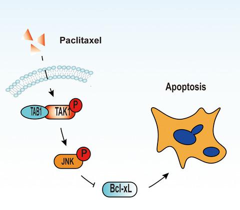
Paclitaxel (PTX) has previously been used to treat tumours of various tissue origins, such as lung, breast, ovarian, prostate cancers and leukemia. PTX‐induced apoptosis is associated with p38 mitogen‐activated protein kinase (p38 MAPK), extracellular signal‐regulated kinase (ERK), nuclear factor‐kappa B (NF‐κB) and c‐Jun N‐terminal kinase or stress‐activated protein kinase (JNK/ SAPK) pathways. Transforming growth factor‐beta‐activated kinase 1 (TAK1) and TAK1‐binding protein 1 (TAB1) play an important role in cell apoptosis through the p38, ERK, NF‐κB and JNK signal transduction pathways. To investigate the role of TAK1 in PTX‐induced cell apoptosis, we treated HEK293 and 8305C cells with 0–20 µM PTX for 6, 12 or 24 h. To investigate whether TAK1 can cooperate with PTX for cancer treatment, we transfected cells with TAK1, TAB1 or control plasmid and treated them with PTX (3–10 µM) for 9–24 h. Apoptosis rates were analysed by flow cytometry (Annexin V/PI). Endogenous TAK1 and TAB1, caspase‐7 cleavage, poly ADP‐ribose polymerase (PARP) cleavage, Bcl‐xL level, phospho‐p44/42, phospho‐JNK and phospho‐p38 were detected by western blot. We show that in HEK293 and 8305C cells, PTX enhanced the endogenous TAK1/TAB1 level and induced cell apoptosis in a dose‐ and time‐dependent manner. Upon TAK1 overexpression in HEK293 cells treated with PTX, apoptosis rate, JNK phosphorylation and PARP cleavage increased contrary to heat‐shocked or untreated cells. CRISPR editing of the tak1 gene upon PTX treatment resulted in lower phospho‐JNK and PARP cleavage levels than in cells transfected with the control or the TAK1‐ or TAB1 + TAK1‐containing plasmids. TAK1‐K63A could not induce JNK phosphorylation or PARP cleavage. We conclude that PTX induces HEK293 and 8305C cell apoptosis through the TAK1–JNK activation pathway, potentially highlighting TAK1’s role in chemosensitivity.
中文翻译:

紫杉醇通过 TAK1-JNK 激活途径诱导细胞凋亡。
紫杉醇 (PTX) 以前曾用于治疗各种组织来源的肿瘤,例如肺癌、乳腺癌、卵巢癌、前列腺癌和白血病。PTX 诱导的细胞凋亡与 p38 丝裂原活化蛋白激酶 (p38 MAPK)、细胞外信号调节激酶 (ERK)、核因子-κB (NF-κB) 和 c-Jun N 端激酶或应激活化蛋白有关激酶 (JNK/SAPK) 通路。转化生长因子-β 活化激酶 1 (TAK1) 和 TAK1 结合蛋白 1 (TAB1) 通过 p38、ERK、NF-κB 和 JNK 信号转导途径在细胞凋亡中发挥重要作用。为了研究 TAK1 在 PTX 诱导的细胞凋亡中的作用,我们用 0–20 µM处理 HEK293 和 8305C 细胞PTX 6、12 或 24 小时。为了研究 TAK1 是否可以与 PTX 合作进行癌症治疗,我们用 TAK1、TAB1 或对照质粒转染细胞,并用 PTX(3-10 µM)处理 9-24 小时。通过流式细胞术(Annexin V/PI)分析细胞凋亡率。通过蛋白质印迹检测内源性 TAK1 和 TAB1、caspase-7 裂解、聚 ADP-核糖聚合酶 (PARP) 裂解、Bcl-xL 水平、磷酸-p44/42、磷酸-JNK 和磷酸-p38。我们表明,在 HEK293 和 8305C 细胞中,PTX 以剂量和时间依赖性方式增强内源性 TAK1/TAB1 水平并诱导细胞凋亡。在用 PTX 处理的 HEK293 细胞中 TAK1 过表达后,与热休克或未处理的细胞相反,细胞凋亡率、JNK 磷酸化和 PARP 裂解增加。tak1 的CRISPR 编辑与用对照或含有 TAK1 或 TAB1 + TAK1 的质粒转染的细胞相比,PTX 处理后的 PTX 基因导致较低的磷酸化 JNK 和 PARP 切割水平。TAK1-K63A 不能诱导 JNK 磷酸化或 PARP 裂解。我们得出结论,PTX 通过 TAK1-JNK 激活途径诱导 HEK293 和 8305C 细胞凋亡,这可能突出了 TAK1 在化学敏感性中的作用。
更新日期:2020-07-31
中文翻译:
紫杉醇通过 TAK1-JNK 激活途径诱导细胞凋亡。
紫杉醇 (PTX) 以前曾用于治疗各种组织来源的肿瘤,例如肺癌、乳腺癌、卵巢癌、前列腺癌和白血病。PTX 诱导的细胞凋亡与 p38 丝裂原活化蛋白激酶 (p38 MAPK)、细胞外信号调节激酶 (ERK)、核因子-κB (NF-κB) 和 c-Jun N 端激酶或应激活化蛋白有关激酶 (JNK/SAPK) 通路。转化生长因子-β 活化激酶 1 (TAK1) 和 TAK1 结合蛋白 1 (TAB1) 通过 p38、ERK、NF-κB 和 JNK 信号转导途径在细胞凋亡中发挥重要作用。为了研究 TAK1 在 PTX 诱导的细胞凋亡中的作用,我们用 0–20 µM处理 HEK293 和 8305C 细胞PTX 6、12 或 24 小时。为了研究 TAK1 是否可以与 PTX 合作进行癌症治疗,我们用 TAK1、TAB1 或对照质粒转染细胞,并用 PTX(3-10 µM)处理 9-24 小时。通过流式细胞术(Annexin V/PI)分析细胞凋亡率。通过蛋白质印迹检测内源性 TAK1 和 TAB1、caspase-7 裂解、聚 ADP-核糖聚合酶 (PARP) 裂解、Bcl-xL 水平、磷酸-p44/42、磷酸-JNK 和磷酸-p38。我们表明,在 HEK293 和 8305C 细胞中,PTX 以剂量和时间依赖性方式增强内源性 TAK1/TAB1 水平并诱导细胞凋亡。在用 PTX 处理的 HEK293 细胞中 TAK1 过表达后,与热休克或未处理的细胞相反,细胞凋亡率、JNK 磷酸化和 PARP 裂解增加。tak1 的CRISPR 编辑与用对照或含有 TAK1 或 TAB1 + TAK1 的质粒转染的细胞相比,PTX 处理后的 PTX 基因导致较低的磷酸化 JNK 和 PARP 切割水平。TAK1-K63A 不能诱导 JNK 磷酸化或 PARP 裂解。我们得出结论,PTX 通过 TAK1-JNK 激活途径诱导 HEK293 和 8305C 细胞凋亡,这可能突出了 TAK1 在化学敏感性中的作用。


 京公网安备 11010802027423号