Journal of Neuroscience Methods ( IF 2.7 ) Pub Date : 2020-06-27 , DOI: 10.1016/j.jneumeth.2020.108828 Jian Wang 1 , Hu Cheng 2 , Sharlene D Newman 2
|
Background
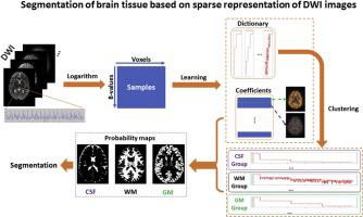
Brain tissue segmentation plays an important role in biomedical research and clinical applications. Traditional segmentation is performed on T1-weighted and/or T2-weighted MRI images. Recently, brain segmentation based on diffusion weighted imaging (DWI) has attracted research interest due to its advantage in diffusion MRI image processing and anatomically-constrained tractography.
New method
We propose a fully automated brain segmentation method based on sparse representation of DWI signals and applied it on nine healthy subjects of Human Connectome Project aged 25–35 years. Learning a dictionary from DWI signals of each subject, brain voxels are classified into gray matter (GM), white matter (WM), and cerebrospinal fluid (CSF) according to their sparse representation of clustered dictionary atoms, achieving good agreement with the segmentation on T1-weighted images using SPM12, as assessed by the DICE score.
Results
The average DICE score for all nine subjects was 0.814 for CSF, 0.850 for GM, and 0.890 for WM. The proposed method is very fast and robust for a wide range of sparse coding parameter selection. It also works well on DWI data with less number of shells or gradient directions.
Comparison with existing methods
On average, our segmentation results are superior to previous methods for all three brain tissue classes in terms of DICE scores.
Conclusion
The proposed method demonstrates the feasibility of segmenting the brain solely based on the tissue response to diffusion encoding.
中文翻译:
用于全自动脑组织分割的DWI图像的稀疏表示。
背景
脑组织分割在生物医学研究和临床应用中起着重要作用。传统分割是在T1加权和/或T2加权MRI图像上执行的。近年来,基于弥散加权成像(DWI)的脑分割技术由于其在弥散MRI图像处理和解剖学约束的术中的优势而吸引了研究兴趣。
新方法
我们提出了一种基于DWI信号稀疏表示的全自动大脑分割方法,并将其应用于25-35岁的人类Connectome项目的9位健康受试者。从每个对象的DWI信号中学习字典,根据其簇字典原子的稀疏表示,将大脑体素分为灰质(GM),白质(WM)和脑脊髓液(CSF),与在使用DPM得分评估的使用SPM12的T1加权图像。
结果
所有九名受试者的平均DICE评分CSF为0.814,GM为0.850,WM为0.890。所提出的方法对于广泛的稀疏编码参数选择非常快速且鲁棒。它也适用于壳数或渐变方向较少的DWI数据。
与现有方法的比较
平均而言,就DICE得分而言,我们的分割结果优于所有三个脑组织类别的分割方法。
结论
所提出的方法证明了仅根据组织对扩散编码的响应来分割大脑的可行性。


 京公网安备 11010802027423号