Journal of Photochemistry and Photobiology B: Biology ( IF 5.4 ) Pub Date : 2020-06-26 , DOI: 10.1016/j.jphotobiol.2020.111944 Nasrin Mohajeri 1 , Ebrahim Mostafavi 2 , Nosratollah Zarghami 3
|
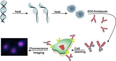
DNA-protein bioconjugation is an appealing target-tracking strategy. The new capability of DNA molecule as a biological nanomaterial endows unique fluorescence and physicochemical properties to be applied in bioimaging. Progression in targeted imaging is contingent on the conjugation of diagnostic nanoparticles to biomolecular signatures, particularly antibody-based ligands. Here, we have reported our recent experience, DNA-dot synthesis and characterization, the covalent conjugation of DNA-dot to goat F(ab')2 IgG and Epidermal Growth Factor Receptor (EGFR) antibodies, [email protected] coupling confirmation, and fluorescent targeted imaging of lung cancer cell line. As a result, the average size of DNA-dot was 4.5–5 nm which was conjugated to amine-rich antibodies with returned PO4−1 groups on the DNA-dot surface via PN bond. The synthetic DNA-dots were conjugated to the goat F(ab')2 IgG and tested for fluorescent detection usability by indirect Dot-blot assay. Also, [email protected] conjugates identified lung cancer cells with 84–92% specificity and 100% sensitivity in five concentrations, associated with 0.0025 to 0.04 g 100 μL−1 DNA-dot. The results demonstrated that bioconjugated DNA-dot can do the diagnosis profiling of molecular biomarkers. Generally, DNA-dot bioconjugation with antibody is implemented within two days and biomarker detection takes one day. Consequently, [email protected] is potentially a toxic-free, swift, and efficient method of antibody labeling that opens up new horizons in fluorescent nanoimaging in the field of cancer cell detection.
中文翻译:
DNA点生物缀合物与抗体的靶向体外癌细胞荧光成像的可行性和可用性。
DNA-蛋白质生物共轭是一种有吸引力的目标跟踪策略。DNA分子作为生物纳米材料的新功能赋予其独特的荧光和理化特性,可用于生物成像。靶向成像的进展取决于诊断性纳米颗粒与生物分子标记(尤其是基于抗体的配体)的缀合。在这里,我们报告了我们最近的经验,DNA点的合成和表征,DNA点与山羊F(ab')2 IgG和表皮生长因子受体(EGFR)抗体的共价缀合,[电子邮件保护的]偶联确认,以及肺癌细胞系的荧光靶向成像。结果,DNA点的平均大小为4.5-5 nm,该点与具有PO 4 -1返回的富含胺的抗体缀合通过PN键在DNA点表面上的基团。将合成的DNA点缀合到山羊F(ab')2 IgG,并通过间接Dot印迹分析测试荧光检测的可用性。同样,[电子邮件保护的]缀合物鉴定出的肺癌细胞在五个浓度下具有84–92%的特异性和100%的敏感性,与0.0025至0.04 g 100μL -1相关DNA点。结果表明,生物缀合的DNA点可以做分子生物标志物的诊断分析。通常,与抗体的DNA点生物缀合在两天内完成,而生物标记物检测则需要一天。因此,[电子邮件保护]可能是一种无毒,快速且有效的抗体标记方法,为癌细胞检测领域的荧光纳米成像开辟了新领域。


 京公网安备 11010802027423号