Acta Tropica ( IF 2.1 ) Pub Date : 2020-06-25 , DOI: 10.1016/j.actatropica.2020.105599 Lana M El-Amin 1 , K E Khalid 2 , Ayman A El-Badry 3
|
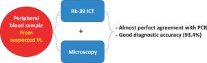
Visceral Leishmaniasis (VL), a life-threating disease in Sudan and Eastern Africa, is usually diagnosed by a painful and invasive tissue aspirate microscopy. This study assessed the diagnostic effectiveness of buffy coat (BC) microscopy and the rK39 immunoassay test separately and combined as an easy non-invasive method applied to peripheral blood sample for field diagnosis of VL. 151 VL suspected patients were recruited from tertiary rural hospitals in Bazura, Gedaref state, from 2014-2015. All patients were tested for VL using rK39 ICT and microscopy smears from LN aspirate and BC in addition to PCR from BC as a reference standard test. Both BC and LN aspirate microscopy showed perfect specificity (100%) with false negative results, while the majority of true positives (81%) had a low-parasite burden. ICT showed almost perfect agreement but limited by its poor specificity. Each of these three tests is inadequate as a consistent single diagnostic tool and should be replaced by PCR in routine practice. Combining the results of risk-free BC microscopy and rk39 ICT, using peripheral blood sample, improved VL diagnosis with almost perfect agreement and 93.4% accuracy. Our findings indicate that combined BC microscopy and ICT are accurate, simple and easy point-of-care VL diagnostic tools in community and rural hospitals that can replace or reduce the use of invasive tissue aspirates microscopy, when PCR is unavailable. This is particularly of value in endemic rural areas, decreasing the delay in final diagnosis and preventing deaths caused by VL.
中文翻译:
无风险即时护理内脏利什曼病的诊断:结合血沉棕黄层显微镜和免疫测定法在苏丹的三级农村医院进行。
内脏利什曼病(VL)是苏丹和东非致命的疾病,通常通过痛苦的浸润性组织抽吸显微镜检查来诊断。这项研究分别评估了血沉棕黄层(BC)显微镜和rK39免疫测定测试的诊断效果,并结合了一种简便,无创的方法应用于外周血样本进行VL现场诊断。2014年至2015年,从Gedaref州Bazura的三级农村医院招募了151例VL疑似患者。除了使用BC的PCR作为参考标准测试外,还使用rK39 ICT和LN吸出液和BC的显微镜涂片对所有患者进行了VL检测。BC和LN抽吸显微镜均显示出完美的特异性(100%),假阴性结果,而大多数真实阳性(81%)的寄生虫负担低。ICT表现出几乎完美的协议,但由于其特异性差而受到限制。这三个测试中的每一个都不足以作为一致的单一诊断工具,在常规实践中应以PCR代替。将无风险的BC显微镜检查结果和rk39 ICT的结果结合在一起,使用外周血样本,以几乎完美的一致性和93.4%的准确度改善了VL诊断。我们的研究结果表明,在社区和乡村医院中,结合使用BC显微镜和ICT是准确,简单,方便的即时医疗VL诊断工具,当无法使用PCR时,可以替代或减少使用侵入性组织抽吸显微镜。这在农村地区很有价值,减少了最终诊断的延迟,并防止了由VL引起的死亡。这三个测试中的每一个都不足以作为一致的单一诊断工具,在常规实践中应以PCR代替。将无风险的BC显微镜检查结果和rk39 ICT的结果结合在一起,使用外周血样本,以几乎完美的一致性和93.4%的准确度改善了VL诊断。我们的研究结果表明,在社区和乡村医院中,结合使用BC显微镜和ICT是准确,简单,方便的即时医疗VL诊断工具,当无法使用PCR时,可以代替或减少使用侵入性组织抽吸显微镜。这在农村地区很有价值,减少了最终诊断的延迟,并防止了由VL引起的死亡。这三个测试中的每一个都不足以作为一致的单一诊断工具,在常规实践中应以PCR代替。将无风险的BC显微镜检查结果和rk39 ICT的结果结合在一起,使用外周血样本,以几乎完美的一致性和93.4%的准确度改善了VL诊断。我们的研究结果表明,在社区和乡村医院中,结合使用BC显微镜和ICT是准确,简单,方便的即时医疗VL诊断工具,当无法使用PCR时,可以代替或减少使用侵入性组织抽吸显微镜。这在农村地区很有价值,减少了最终诊断的延迟,并防止了由VL引起的死亡。改善了VL诊断,具有几乎完美的一致性和93.4%的准确度。我们的研究结果表明,在社区和乡村医院中,结合使用BC显微镜和ICT是准确,简单,方便的即时医疗VL诊断工具,当无法使用PCR时,可以替代或减少使用侵入性组织抽吸显微镜。这在农村地区很有价值,减少了最终诊断的延迟,并防止了由VL引起的死亡。改善了VL诊断,具有几乎完美的一致性和93.4%的准确性。我们的研究结果表明,在社区和乡村医院中,结合使用BC显微镜和ICT是准确,简单,方便的即时医疗VL诊断工具,当无法使用PCR时,可以代替或减少使用侵入性组织抽吸显微镜。这在农村地区很有价值,减少了最终诊断的延迟,并防止了由VL引起的死亡。


 京公网安备 11010802027423号