当前位置:
X-MOL 学术
›
J. Morphol.
›
论文详情
Our official English website, www.x-mol.net, welcomes your
feedback! (Note: you will need to create a separate account there.)
The neuroactive substances and associated muscle system in Rhipidocotyle campanula (Digenea, Bucephalidae) from the intestine of the pike Esox lucius
Journal of Morphology ( IF 1.5 ) Pub Date : 2020-06-23 , DOI: 10.1002/jmor.21230 Natalia Kreshchenko 1 , Nadezhda Terenina 2 , Darya Nefedova 2 , Natalia Mochalova 2 , Ekaterina Voropaeva 2, 3 , Sergey Movsesyan 2
Journal of Morphology ( IF 1.5 ) Pub Date : 2020-06-23 , DOI: 10.1002/jmor.21230 Natalia Kreshchenko 1 , Nadezhda Terenina 2 , Darya Nefedova 2 , Natalia Mochalova 2 , Ekaterina Voropaeva 2, 3 , Sergey Movsesyan 2
Affiliation
|
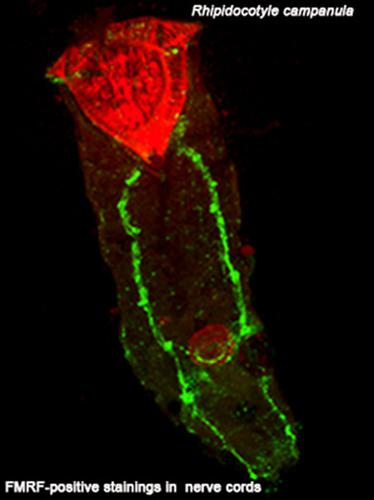
We report about the muscular system and the serotonergic and FMRFamidergic components of the nervous system of the Bucephalidae trematode, Rhipidocotyle campanula, an intestinal parasite of the pike. We use immunocytochemical methods and confocal scanning laser microscopy (CLSM). The musculature is identified by histochemical staining with fluorescently labeled phalloidin. The body wall musculature of R. campanula contains three layers of muscle fibres – the outer thin circular, intermediate longitudinal and inner diagonal muscle fibres running in two opposite directions. The digestive system of R. campanula possess of a well‐developed musculature: radial, longitudinal and circular muscle elements are detected in the pharynx, circular and longitudinal muscle filaments seen in the oesophagus, and longitudinal and the circular muscle fibres were found in the intestinal wall. Specific staining indicating the presence of actin muscle filaments occurs in the cirrus sac localized in the posterior body region. The frontal region of anterior attachment organ, the rhynchus, in R. campanula is represented by radial muscle fibres. The posterior part of the rhynchus comprise of radial muscles forming the organ's wall, and several strong longitudinal muscle bundles. Serotonergic and FMRFamidergic structures are detected in the central and peripheral compartments of the nervous system of R. campanula, that is, in the paired brain ganglia, the brain commissure, the longitudinal nerve cords, and connective nerve commissures. The innervations of the rhynchus, pharynx, oesophagus and distal regions of the reproductive system by the serotonergic and FMRFamidergic nervous elements are revealed. We compare our findings obtained on R. campanula with related data for other trematodes.
中文翻译:

来自梭鱼肠道的风铃草 (Digenea, Bucephalidae) 中的神经活性物质和相关肌肉系统
我们报告了布氏吸虫、Rhipidocotyle campanula(一种梭鱼的肠道寄生虫)的神经系统的肌肉系统和血清素能和 FMRFamidergic 成分。我们使用免疫细胞化学方法和共聚焦扫描激光显微镜 (CLSM)。通过用荧光标记的鬼笔环肽进行组织化学染色来鉴定肌肉组织。R. campanula 的体壁肌肉组织包含三层肌纤维——外薄圆形肌纤维、中间纵向肌纤维和向两个相反方向延伸的内斜肌纤维。风铃草的消化系统具有发达的肌肉组织:在咽部检测到径向、纵向和圆形肌肉成分,在食道中可见圆形和纵向肌丝,肠壁可见纵向和环状肌纤维。表明存在肌动蛋白肌丝的特定染色发生在位于身体后部区域的卷云囊中。R.campanula 中前附着器官的额叶区域,rhynchus,由径向肌纤维代表。rhynchus 的后部由形成器官壁的径向肌肉和几个强大的纵向肌肉束组成。在风铃草神经系统的中枢和外周区室中检测到血清素能和 FMRFamidergic 结构,即在成对的脑神经节、脑连合、纵向神经索和结缔神经连合中。鼻、咽的神经支配,食道和生殖系统的远端区域由血清素能和 FMRFamidergic 神经元件显示出来。我们将我们在风铃草上获得的发现与其他吸虫的相关数据进行了比较。
更新日期:2020-06-23
中文翻译:
来自梭鱼肠道的风铃草 (Digenea, Bucephalidae) 中的神经活性物质和相关肌肉系统
我们报告了布氏吸虫、Rhipidocotyle campanula(一种梭鱼的肠道寄生虫)的神经系统的肌肉系统和血清素能和 FMRFamidergic 成分。我们使用免疫细胞化学方法和共聚焦扫描激光显微镜 (CLSM)。通过用荧光标记的鬼笔环肽进行组织化学染色来鉴定肌肉组织。R. campanula 的体壁肌肉组织包含三层肌纤维——外薄圆形肌纤维、中间纵向肌纤维和向两个相反方向延伸的内斜肌纤维。风铃草的消化系统具有发达的肌肉组织:在咽部检测到径向、纵向和圆形肌肉成分,在食道中可见圆形和纵向肌丝,肠壁可见纵向和环状肌纤维。表明存在肌动蛋白肌丝的特定染色发生在位于身体后部区域的卷云囊中。R.campanula 中前附着器官的额叶区域,rhynchus,由径向肌纤维代表。rhynchus 的后部由形成器官壁的径向肌肉和几个强大的纵向肌肉束组成。在风铃草神经系统的中枢和外周区室中检测到血清素能和 FMRFamidergic 结构,即在成对的脑神经节、脑连合、纵向神经索和结缔神经连合中。鼻、咽的神经支配,食道和生殖系统的远端区域由血清素能和 FMRFamidergic 神经元件显示出来。我们将我们在风铃草上获得的发现与其他吸虫的相关数据进行了比较。


 京公网安备 11010802027423号