Acta Neurochirurgica ( IF 1.9 ) Pub Date : 2020-06-23 , DOI: 10.1007/s00701-020-04452-0 Miki Katzir 1 , Nguyen Hoang 1 , Eric Bourekas 2 , Ricardo Carrau 3 , Ehud Mendel 1
|
Background
Metastatic cervical spine disease can cause compression fractures, cervical spine instability, and pain. Vertebroplasty can stabilize a fracture, reduce the pain associated with a compression fracture, prevent or stop the progression of a fracture, thus avoiding cervical spine fixation, and decreased mobility. Transoral C2 vertebroplasty is less invasive than open fusion surgery, but it poses its own risk of infection and cement leak in this highly sensitive area.
Methods
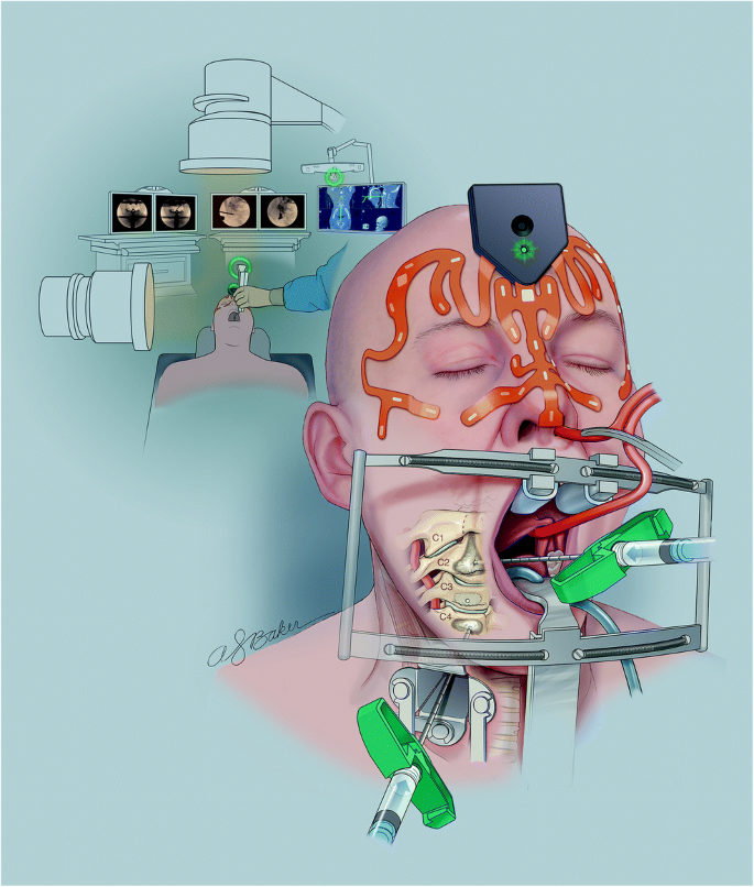
The image guidance setup consisted of the Stryker NAV3i navigation system, Stryker CranialMask tracker, and the CranialMap 3.0 software combined with biplanar fluoroscopy.
Results
The patient’s neck pain has completely resolved immediately after the surgery. There were no complications.
Conclusion
Quality of life preservation is paramount in the management of metastatic spine disease. Vertebroplasty of osteolytic lesions can both relieve pain and restore stability, thus avoiding permanent stiff cervical collar, halo vest, or upfront occipitocervical fusion. With the increasing availability of surgical navigation systems, its use combined with biplanar fluoroscopy for performing transoral C2 vertebroplasty seems to be an adequate treatment in selected cases for pain relief, stabilization, and maintaining quality of life in the complex cancer population with C2 pathological fractures. The article describes as well vertebroplasty of the subaxial spine through a conventional anterior approach which again seems to be adequate in the treatment of spinal pathological fractures.
中文翻译:
立体定向CT图像引导和双平面荧光检查用于经口C2椎体成形术和直接前外侧亚轴椎体成形术:关于进入轴突和亚轴脊柱的外科手术技术说明。
背景
转移性颈椎疾病可导致压迫性骨折,颈椎不稳定和疼痛。椎体成形术可以稳定骨折,减轻与压缩性骨折相关的疼痛,防止或阻止骨折的进展,从而避免颈椎固定和活动性降低。经口C2椎体成形术比开放融合术的侵袭性小,但在这个高度敏感的区域内,存在着自身感染和水泥渗漏的风险。
方法
图像导航设置由Stryker NAV3i导航系统,Stryker CranialMask跟踪器以及结合了双平面荧光检查的CranialMap 3.0软件组成。
结果
手术后,患者的颈部疼痛已完全消除。没有并发症。
结论
在转移性脊柱疾病的治疗中,维持生命的质量至关重要。溶骨性病变的椎体成形术既可以缓解疼痛又可以恢复稳定性,从而避免永久性僵硬的颈托,光晕背心或前枕骨融合。随着外科手术导航系统可用性的提高,结合使用双平面透视检查进行经口C2椎体成形术似乎是某些病例的适当治疗,以减轻,稳定和维持具有C2病理性骨折的复杂癌症人群的生活质量。该文章还通过常规的前路方法描述了亚轴脊柱椎体成形术,这似乎也足以治疗脊柱病理性骨折。


 京公网安备 11010802027423号