当前位置:
X-MOL 学术
›
Microsc. Res. Tech.
›
论文详情
Our official English website, www.x-mol.net, welcomes your feedback! (Note: you will need to create a separate account there.)
Sample preparation and microscopical investigation techniques for metal and ceramics containing graphite and graphene-like layered particles.
Microscopy Research and Technique ( IF 2.5 ) Pub Date : 2020-06-20 , DOI: 10.1002/jemt.23521 Sinem Baskut 1 , Servet Turan 1
Microscopy Research and Technique ( IF 2.5 ) Pub Date : 2020-06-20 , DOI: 10.1002/jemt.23521 Sinem Baskut 1 , Servet Turan 1
Affiliation
|
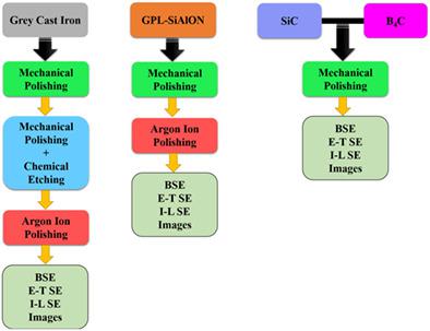
Scanning electron microscopy (SEM) techniques are widely used in microstructural investigations of materials since it can provide surface morphology, topography, and chemical information. However, it is important to use correct imaging and sample preparation techniques to reveal the microstructures of materials composed of components with different polishing characteristics such as grey cast iron, graphene platelets (GPLs)‐added SiAlON composite, SiC and B4C ceramics containing graphite or graphene‐like layered particles. In this study, all microstructural details of gray cast iron were successfully revealed by using argon ion beam milling as an alternative to the standard sample preparation method for cast irons, that is, mechanical polishing followed by chemical etching. The in‐lens secondary electron (I‐L‐SE) image was clearly displayed on the surface details of the graphites that could not be revealed by backscattered electron (BSE) and Everhart–Thornley secondary electron (E‐T SE) images. Mechanical polishing leads to pull‐out of GPLs from SiAlON surface, whereas argon ion beam milling preserved the GPLs and resulted in smooth surface. Grain and grain boundaries of polycrystalline SiC and B4C were easily revealed by using I‐L SE image in the SEM after only mechanical polishing without any etching process. While the BSE and E‐T SE images did not clearly show the residual graphites in the microstructure, their distribution in the B4C matrix was fully revealed in the I‐L SE image.
中文翻译:

包含石墨和类石墨烯层状颗粒的金属和陶瓷的样品制备和显微研究技术。
扫描电子显微镜(SEM)技术可广泛用于材料的微结构研究,因为它可以提供表面形态,形貌和化学信息。但是,重要的是要使用正确的成像和样品制备技术来揭示由具有不同抛光特性的组件(例如灰铸铁,石墨烯薄片(GPL)添加的SiAlON复合材料,SiC和B 4)组成的材料的微观结构。包含石墨或类石墨烯层状颗粒的C陶瓷。在这项研究中,通过使用氩离子束铣削替代铸铁的标准样品制备方法,即机械抛光后进行化学蚀刻,成功地揭示了灰口铸铁的所有微观结构细节。透镜内二次电子(IL-SE)图像清晰地显示在石墨的表面细节上,而反向散射电子(BSE)和Everhart-Thornley二次电子(ETSE)图像无法显示这些细节。机械抛光导致GPL从SiAlON表面拔出,而氩离子束铣削则保留了GPL并产生了光滑的表面。多晶SiC和B 4的晶粒和晶界仅需机械抛光,无需任何蚀刻工艺,即可在SEM中使用I-L SE图像轻松显示C。虽然BSE和E‐T SE图像没有清晰地显示出微观结构中的残留石墨,但它们在B 4 C基质中的分布在I‐L SE图像中得到了充分揭示。
更新日期:2020-06-20
中文翻译:
包含石墨和类石墨烯层状颗粒的金属和陶瓷的样品制备和显微研究技术。
扫描电子显微镜(SEM)技术可广泛用于材料的微结构研究,因为它可以提供表面形态,形貌和化学信息。但是,重要的是要使用正确的成像和样品制备技术来揭示由具有不同抛光特性的组件(例如灰铸铁,石墨烯薄片(GPL)添加的SiAlON复合材料,SiC和B 4)组成的材料的微观结构。包含石墨或类石墨烯层状颗粒的C陶瓷。在这项研究中,通过使用氩离子束铣削替代铸铁的标准样品制备方法,即机械抛光后进行化学蚀刻,成功地揭示了灰口铸铁的所有微观结构细节。透镜内二次电子(IL-SE)图像清晰地显示在石墨的表面细节上,而反向散射电子(BSE)和Everhart-Thornley二次电子(ETSE)图像无法显示这些细节。机械抛光导致GPL从SiAlON表面拔出,而氩离子束铣削则保留了GPL并产生了光滑的表面。多晶SiC和B 4的晶粒和晶界仅需机械抛光,无需任何蚀刻工艺,即可在SEM中使用I-L SE图像轻松显示C。虽然BSE和E‐T SE图像没有清晰地显示出微观结构中的残留石墨,但它们在B 4 C基质中的分布在I‐L SE图像中得到了充分揭示。


 京公网安备 11010802027423号