iScience ( IF 4.6 ) Pub Date : 2020-06-20 , DOI: 10.1016/j.isci.2020.101290 Georg Kislinger 1 , Helmut Gnägi 2 , Martin Kerschensteiner 3 , Mikael Simons 1 , Thomas Misgeld 1 , Martina Schifferer 4
|
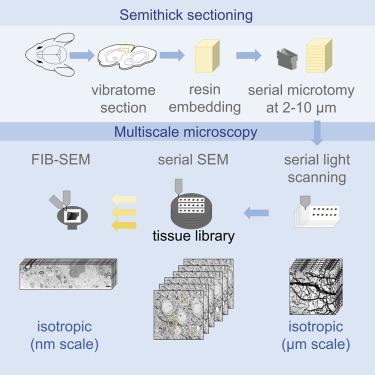
Volume electron microscopy enables the ultrastructural analysis of biological tissue. Currently, the techniques involving ultramicrotomy (ATUM, ssTEM) allow large fields of view but afford only limited z-resolution, whereas ion beam-milling approaches (FIB-SEM) yield isotropic voxels but are restricted in volume size. Now we present a hybrid method, named ATUM-FIB, which combines the advantages of both approaches. ATUM-FIB is based on serial sectioning of tissue into “semithick” (2–10 μm) sections collected onto tape. Serial light and electron microscopy allows the identification of regions of interest that are then directly accessible for targeted FIB-SEM. The set of semithick sections thus represents a tissue “library” which provides three-dimensional context information that can be probed “on demand” by local high-resolution analysis. We demonstrate the potential of this technique to reveal the ultrastructure of rare but pathologically important events by identifying microglia contact sites with amyloid plaques in a mouse model of familial Alzheimer's disease.
中文翻译:
多尺度 ATUM-FIB 显微镜能够以各向同性分辨率进行有针对性的超微结构分析。
体积电子显微镜能够对生物组织进行超微结构分析。目前,涉及超薄切片技术 (ATUM, ssTEM) 的技术允许大视野但仅提供有限的 z 分辨率,而离子束铣削方法 (FIB-SEM) 产生各向同性体素但体积大小受到限制。现在我们提出一种混合方法,称为 ATUM-FIB,它结合了两种方法的优点。ATUM-FIB 基于将组织连续切片成“半厚”(2-10 μm)切片,收集到磁带上。串行光学和电子显微镜可以识别感兴趣的区域,然后可以直接访问目标 FIB-SEM。因此,这组半厚切片代表了一个组织“库”,它提供了可以通过局部高分辨率分析“按需”探测的 3D 上下文信息。


 京公网安备 11010802027423号