Artificial Intelligence in Medicine ( IF 6.1 ) Pub Date : 2020-06-20 , DOI: 10.1016/j.artmed.2020.101911 Wei-Kai Lee , Chih-Chun Wu , Cheng-Chia Lee , Chia-Feng Lu , Huai-Che Yang , Tzu-Hsuan Huang , Chun-Yi Lin , Wen-Yuh Chung , Po-Shan Wang , Hsiu-Mei Wu , Wan-Yuo Guo , Yu-Te Wu
|
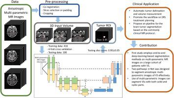
Manual delineation of vestibular schwannoma (VS) by magnetic resonance (MR) imaging is required for diagnosis, radiosurgery dose planning, and follow-up tumor volume measurement. A rapid and objective automatic segmentation method is required, but problems have been encountered due to the low through-plane resolution of standard VS MR scan protocols and because some patients have non-homogeneous cystic areas within their tumors. In this study, we retrospectively collected multi-parametric MR images from 516 patients with VS; these were extracted from the Gamma Knife radiosurgery planning system and consisted of T1-weighted (T1W), T2-weighted (T2W), and T1W with contrast (T1W + C) images. We developed an end-to-end deep-learning-based method via an automatic preprocessing pipeline. A two-pathway U-Net model involving two sizes of convolution kernel (i.e., 3 × 3 × 1 and 1 × 1 × 3) was used to extract the in-plane and through-plane features of the anisotropic MR images. A single-pathway model that adopted the same architecture as the two-pathway model, but used a kernel size of 3 × 3 × 3, was also developed for comparison purposes. In addition, we used multi-parametric MR images with different image contrasts as the model training input in order to effectively segment tumors with solid as well as cystic parts. The results of the automatic segmentation demonstrated that (1) the two-pathway model outperformed single-pathway model in terms of dice scores (0.90 ± 0.05 versus 0.87 ± 0.07); both of them having been trained using the T1W, T1W + C and T2W anisotropic MR images, (2) the optimal single-parametric two-pathway model (dice score: 0.88 ± 0.06) was then trained using the T1W + C images, and (3) the two-pathway models trained using bi-parametric (T1W + C and T2W) and tri-parametric (T1W, T2W, and T1W + C) images outperformed the model trained using the single-parametric (T1W + C) images (dice scores: 0.89 ± 0.05 and 0.90 ± 0.05, respectively, larger than 0.88 ± 0.06) because it showed improved segmentation of the non-homogeneous parts of the tumors. The proposed two-pathway U-Net model outperformed the single-pathway U-Net model when segmenting VS using anisotropic MR images. The multi-parametric models effectively improved on the defective segmentation obtained using the single-parametric models by separating the non-homogeneous tumors into their solid and cystic parts.
中文翻译:
将多参数 MR 图像分析结合到卷积神经网络中:前庭神经鞘瘤治疗计划的精确目标描绘。
诊断、放射外科剂量规划和随访肿瘤体积测量需要通过磁共振 (MR) 成像手动描绘前庭神经鞘瘤 (VS)。需要一种快速、客观的自动分割方法,但由于标准 VS MR 扫描协议的平面分辨率低以及一些患者的肿瘤内具有非均匀囊性区域,因此遇到了问题。在这项研究中,我们回顾性收集了 516 名 VS 患者的多参数 MR 图像;这些图像是从伽玛刀放射外科计划系统中提取的,由 T1 加权 (T1W)、T2 加权 (T2W) 和具有对比度 (T1W + C) 的 T1W 图像组成。我们通过自动预处理管道开发了一种基于端到端深度学习的方法。使用包含两种尺寸的卷积核(即 3 × 3 × 1 和 1 × 1 × 3)的双通路 U-Net 模型来提取各向异性 MR 图像的面内和面内特征。还开发了一个单通路模型,该模型采用与双通路模型相同的架构,但使用 3 × 3 × 3 的内核大小,用于比较目的。此外,我们使用具有不同图像对比度的多参数 MR 图像作为模型训练输入,以有效地分割具有实性和囊性部分的肿瘤。自动分割的结果表明:(1)双通路模型在骰子得分方面优于单通路模型(0.90±0.05 vs 0.87±0.07);他们都使用 T1W、T1W + C 和 T2W 各向异性 MR 图像进行了训练,(2) 然后使用 T1W + C 图像训练最佳单参数双通路模型(骰子分数:0.88 ± 0.06),以及 (3) 使用双参数(T1W + C 和 T2W)训练的双通路模型) 和三参数 (T1W、T2W 和 T1W + C) 图像优于使用单参数 (T1W + C) 图像训练的模型(骰子分数:分别为 0.89 ± 0.05 和 0.90 ± 0.05,大于 0.88 ± 0.06 ) 因为它显示了对肿瘤非均质部分的改进分割。当使用各向异性 MR 图像分割 VS 时,提出的双通路 U-Net 模型优于单通路 U-Net 模型。多参数模型通过将非均质肿瘤分成实性和囊性部分,有效地改进了使用单参数模型获得的缺陷分割。06) 然后使用 T1W + C 图像进行训练,并且 (3) 使用双参数(T1W + C 和 T2W)和三参数(T1W、T2W 和 T1W + C)图像训练的双通路模型优于使用单参数 (T1W + C) 图像(骰子分数:分别为 0.89 ± 0.05 和 0.90 ± 0.05,大于 0.88 ± 0.06)训练的模型,因为它显示了对肿瘤非均质部分的改进分割。当使用各向异性 MR 图像分割 VS 时,提出的双通路 U-Net 模型优于单通路 U-Net 模型。多参数模型通过将非均质肿瘤分成实性和囊性部分,有效地改进了使用单参数模型获得的缺陷分割。06) 然后使用 T1W + C 图像进行训练,并且 (3) 使用双参数(T1W + C 和 T2W)和三参数(T1W、T2W 和 T1W + C)图像训练的双通路模型优于使用单参数 (T1W + C) 图像(骰子分数:分别为 0.89 ± 0.05 和 0.90 ± 0.05,大于 0.88 ± 0.06)训练的模型,因为它显示了对肿瘤非均质部分的改进分割。当使用各向异性 MR 图像分割 VS 时,提出的双通路 U-Net 模型优于单通路 U-Net 模型。多参数模型通过将非均质肿瘤分成实性和囊性部分,有效地改进了使用单参数模型获得的缺陷分割。(3) 使用双参数(T1W + C 和 T2W)和三参数(T1W、T2W 和 T1W + C)图像训练的双通路模型优于使用单参数 (T1W + C) 训练的模型图像(骰子分数:分别为 0.89 ± 0.05 和 0.90 ± 0.05,大于 0.88 ± 0.06),因为它显示了对肿瘤非均质部分的改进分割。在使用各向异性 MR 图像分割 VS 时,所提出的双通路 U-Net 模型优于单通路 U-Net 模型。多参数模型通过将非均质肿瘤分为实性和囊性部分,有效地改进了使用单参数模型获得的缺陷分割。(3) 使用双参数(T1W + C 和 T2W)和三参数(T1W、T2W 和 T1W + C)图像训练的双通路模型优于使用单参数 (T1W + C) 训练的模型图像(骰子分数:分别为 0.89 ± 0.05 和 0.90 ± 0.05,大于 0.88 ± 0.06),因为它显示了对肿瘤非均质部分的改进分割。在使用各向异性 MR 图像分割 VS 时,所提出的双通路 U-Net 模型优于单通路 U-Net 模型。多参数模型通过将非均质肿瘤分成实性和囊性部分,有效地改进了使用单参数模型获得的缺陷分割。分别大于 0.88 ± 0.06),因为它显示出对肿瘤非均质部分的改进分割。当使用各向异性 MR 图像分割 VS 时,提出的双通路 U-Net 模型优于单通路 U-Net 模型。多参数模型通过将非均质肿瘤分成实性和囊性部分,有效地改进了使用单参数模型获得的缺陷分割。分别大于 0.88 ± 0.06),因为它显示出对肿瘤非均质部分的改进分割。当使用各向异性 MR 图像分割 VS 时,提出的双通路 U-Net 模型优于单通路 U-Net 模型。多参数模型通过将非均质肿瘤分成实性和囊性部分,有效地改进了使用单参数模型获得的缺陷分割。


 京公网安备 11010802027423号