当前位置:
X-MOL 学术
›
Macromol. Biosci.
›
论文详情
Our official English website, www.x-mol.net, welcomes your
feedback! (Note: you will need to create a separate account there.)
Imaging the In Vivo Degradation of Tissue Engineering Implants by Use of Supramolecular Radiopaque Biomaterials.
Macromolecular Bioscience ( IF 4.4 ) Pub Date : 2020-06-17 , DOI: 10.1002/mabi.202000024 Hanna Talacua 1, 2 , Serge H M Söntjens 3 , Shraddha H Thakkar 4 , Aurelie M A Brizard 5 , Lex A van Herwerden 1 , Aryan Vink 6 , Geert C van Almen 7, 8 , Patricia Y W Dankers 7, 8 , Carlijn V C Bouten 4, 8 , Ricardo P J Budde 9 , Henk M Janssen 3 , Jolanda Kluin 1, 2
Macromolecular Bioscience ( IF 4.4 ) Pub Date : 2020-06-17 , DOI: 10.1002/mabi.202000024 Hanna Talacua 1, 2 , Serge H M Söntjens 3 , Shraddha H Thakkar 4 , Aurelie M A Brizard 5 , Lex A van Herwerden 1 , Aryan Vink 6 , Geert C van Almen 7, 8 , Patricia Y W Dankers 7, 8 , Carlijn V C Bouten 4, 8 , Ricardo P J Budde 9 , Henk M Janssen 3 , Jolanda Kluin 1, 2
Affiliation
|
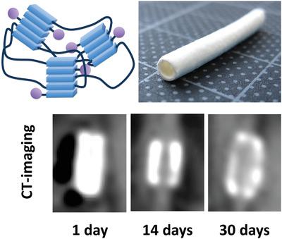
For in situ tissue engineering (TE) applications it is important that implant degradation proceeds in concord with neo‐tissue formation to avoid graft failure. It will therefore be valuable to have an imaging contrast agent (CA) available that can report on the degrading implant. For this purpose, a biodegradable radiopaque biomaterial is presented, modularly composed of a bisurea chain‐extended polycaprolactone (PCL2000‐U4U) elastomer and a novel iodinated bisurea‐modified CA additive (I‐U4U). Supramolecular hydrogen bonding interactions between the components ensure their intimate mixing. Porous implant TE‐grafts are prepared by simply electrospinning a solution containing PCL2000‐U4U and I‐U4U. Rats receive an aortic interposition graft, either composed of only PCL2000‐U4U (control) or of PCL2000‐U4U and I‐U4U (test). The grafts are explanted for analysis at three time points over a 1‐month period. Computed tomography imaging of the test group implants prior to explantation shows a decrease in iodide volume and density over time. Explant analysis also indicates scaffold degradation. (Immuno)histochemistry shows comparable cellular contents and a similar neo‐tissue formation process for test and control group, demonstrating that the CA does not have apparent adverse effects. A supramolecular approach to create solid radiopaque biomaterials can therefore be used to noninvasively monitor the biodegradation of synthetic implants.
中文翻译:

通过使用超分子不透射线生物材料对组织工程植入物的体内降解进行成像。
对于原位组织工程(TE)应用,重要的是植入物的降解与新组织的形成一致进行,以避免移植失败。因此,具有可报告降解植入物的成像造影剂(CA)将会很有价值。为此,我们提出了一种可生物降解的不透射线生物材料,该材料由Bisurea扩链聚己内酯(PCL2000-U4U)弹性体和新型碘化Bisurea改性CA添加剂(I-U4U)组成。组分之间的超分子氢键相互作用确保了它们的紧密混合。多孔植入物TE移植物是通过简单地电纺包含PCL2000-U4U和I-U4U的溶液来制备的。大鼠接受主动脉移植物,由仅PCL2000-U4U(对照)或PCL2000-U4U和I-U4U(测试)组成。在1个月的三个时间点移植移植物以进行分析。植入前测试组植入物的计算机断层扫描成像显示碘化物的体积和密度随时间降低。外植体分析还表明支架降解。(免疫)组织化学显示,测试组和对照组的细胞含量相当,并且新组织的形成过程相似,这表明CA没有明显的不良反应。因此,可以使用超分子方法产生固体不透射线的生物材料,以无创地监测合成植入物的生物降解。外植体分析还表明支架降解。(免疫)组织化学显示,测试组和对照组的细胞含量相当,并且新组织的形成过程相似,这表明CA没有明显的不良反应。因此,可以使用超分子方法产生固体不透射线的生物材料,以无创地监测合成植入物的生物降解。外植体分析还表明支架降解。(免疫)组织化学显示,测试组和对照组的细胞含量相当,并且新组织的形成过程相似,这表明CA没有明显的不良反应。因此,可以使用超分子方法产生固体不透射线的生物材料,以无创地监测合成植入物的生物降解。
更新日期:2020-06-17
中文翻译:
通过使用超分子不透射线生物材料对组织工程植入物的体内降解进行成像。
对于原位组织工程(TE)应用,重要的是植入物的降解与新组织的形成一致进行,以避免移植失败。因此,具有可报告降解植入物的成像造影剂(CA)将会很有价值。为此,我们提出了一种可生物降解的不透射线生物材料,该材料由Bisurea扩链聚己内酯(PCL2000-U4U)弹性体和新型碘化Bisurea改性CA添加剂(I-U4U)组成。组分之间的超分子氢键相互作用确保了它们的紧密混合。多孔植入物TE移植物是通过简单地电纺包含PCL2000-U4U和I-U4U的溶液来制备的。大鼠接受主动脉移植物,由仅PCL2000-U4U(对照)或PCL2000-U4U和I-U4U(测试)组成。在1个月的三个时间点移植移植物以进行分析。植入前测试组植入物的计算机断层扫描成像显示碘化物的体积和密度随时间降低。外植体分析还表明支架降解。(免疫)组织化学显示,测试组和对照组的细胞含量相当,并且新组织的形成过程相似,这表明CA没有明显的不良反应。因此,可以使用超分子方法产生固体不透射线的生物材料,以无创地监测合成植入物的生物降解。外植体分析还表明支架降解。(免疫)组织化学显示,测试组和对照组的细胞含量相当,并且新组织的形成过程相似,这表明CA没有明显的不良反应。因此,可以使用超分子方法产生固体不透射线的生物材料,以无创地监测合成植入物的生物降解。外植体分析还表明支架降解。(免疫)组织化学显示,测试组和对照组的细胞含量相当,并且新组织的形成过程相似,这表明CA没有明显的不良反应。因此,可以使用超分子方法产生固体不透射线的生物材料,以无创地监测合成植入物的生物降解。


 京公网安备 11010802027423号