当前位置:
X-MOL 学术
›
Chem. Sci.
›
论文详情
Our official English website, www.x-mol.net, welcomes your
feedback! (Note: you will need to create a separate account there.)
Templating S100A9 amyloids on Aβ fibrillar surfaces revealed by charge detection mass spectrometry, microscopy, kinetic and microfluidic analyses
Chemical Science ( IF 7.6 ) Pub Date : 2020-06-17 , DOI: 10.1039/c9sc05905a Jonathan Pansieri 1 , Igor A Iashchishyn 1 , Hussein Fakhouri 2 , Lucija Ostojić 1 , Mantas Malisauskas 1 , Greta Musteikyte 3 , Vytautas Smirnovas 3 , Matthias M Schneider 4 , Tom Scheidt 4 , Catherine K Xu 4 , Georg Meisl 4 , Tuomas P J Knowles 4, 5 , Ehud Gazit 1, 6 , Rodolphe Antoine 2 , Ludmilla A Morozova-Roche 1
Chemical Science ( IF 7.6 ) Pub Date : 2020-06-17 , DOI: 10.1039/c9sc05905a Jonathan Pansieri 1 , Igor A Iashchishyn 1 , Hussein Fakhouri 2 , Lucija Ostojić 1 , Mantas Malisauskas 1 , Greta Musteikyte 3 , Vytautas Smirnovas 3 , Matthias M Schneider 4 , Tom Scheidt 4 , Catherine K Xu 4 , Georg Meisl 4 , Tuomas P J Knowles 4, 5 , Ehud Gazit 1, 6 , Rodolphe Antoine 2 , Ludmilla A Morozova-Roche 1
Affiliation
|
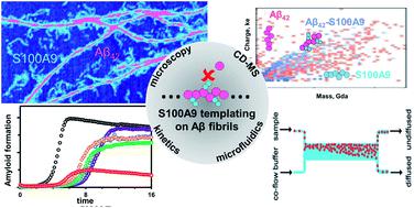
The mechanism of amyloid co-aggregation and its nucleation process are not fully understood in spite of extensive studies. Deciphering the interactions between proinflammatory S100A9 protein and Aβ42 peptide in Alzheimer's disease is fundamental since inflammation plays a central role in the disease onset. Here we use innovative charge detection mass spectrometry (CDMS) together with biophysical techniques to provide mechanistic insight into the co-aggregation process and differentiate amyloid complexes at a single particle level. Combination of mass and charge distributions of amyloids together with reconstruction of the differences between them and detailed microscopy reveals that co-aggregation involves templating of S100A9 fibrils on the surface of Aβ42 amyloids. Kinetic analysis further corroborates that the surfaces available for the Aβ42 secondary nucleation are diminished due to the coating by S100A9 amyloids, while the binding of S100A9 to Aβ42 fibrils is validated by a microfluidic assay. We demonstrate that synergy between CDMS, microscopy, kinetic and microfluidic analyses opens new directions in interdisciplinary research.
中文翻译:

通过电荷检测质谱、显微镜、动力学和微流体分析揭示 Aβ 纤维表面上的 S100A9 淀粉样蛋白模板化
尽管进行了广泛的研究,但淀粉样蛋白共聚集的机制及其成核过程尚未完全了解。破译促炎性 S100A9 蛋白和 Aβ 42肽在阿尔茨海默病中的相互作用至关重要,因为炎症在该病的发病中起着核心作用。在这里,我们使用创新的电荷检测质谱(CDMS)和生物物理技术来提供对共聚集过程的机制洞察,并在单颗粒水平上区分淀粉样蛋白复合物。淀粉样蛋白的质量和电荷分布的组合以及它们之间差异的重建和详细的显微镜揭示了共聚集涉及 Aβ 42淀粉样蛋白表面上的 S100A9 原纤维的模板化。动力学分析进一步证实,由于 S100A9 淀粉样蛋白的涂层,可用于 Aβ 42二次成核的表面减少,而 S100A9 与 Aβ 42原纤维的结合通过微流体测定得到验证。我们证明 CDMS、显微镜、动力学和微流体分析之间的协同作用为跨学科研究开辟了新的方向。
更新日期:2020-07-15
中文翻译:
通过电荷检测质谱、显微镜、动力学和微流体分析揭示 Aβ 纤维表面上的 S100A9 淀粉样蛋白模板化
尽管进行了广泛的研究,但淀粉样蛋白共聚集的机制及其成核过程尚未完全了解。破译促炎性 S100A9 蛋白和 Aβ 42肽在阿尔茨海默病中的相互作用至关重要,因为炎症在该病的发病中起着核心作用。在这里,我们使用创新的电荷检测质谱(CDMS)和生物物理技术来提供对共聚集过程的机制洞察,并在单颗粒水平上区分淀粉样蛋白复合物。淀粉样蛋白的质量和电荷分布的组合以及它们之间差异的重建和详细的显微镜揭示了共聚集涉及 Aβ 42淀粉样蛋白表面上的 S100A9 原纤维的模板化。动力学分析进一步证实,由于 S100A9 淀粉样蛋白的涂层,可用于 Aβ 42二次成核的表面减少,而 S100A9 与 Aβ 42原纤维的结合通过微流体测定得到验证。我们证明 CDMS、显微镜、动力学和微流体分析之间的协同作用为跨学科研究开辟了新的方向。


 京公网安备 11010802027423号