当前位置:
X-MOL 学术
›
NeuroImage
›
论文详情
Our official English website, www.x-mol.net, welcomes your
feedback! (Note: you will need to create a separate account there.)
Cingulate cortex function and multi-modal connectivity mapped using intracranial stimulation
NeuroImage ( IF 4.7 ) Pub Date : 2020-10-01 , DOI: 10.1016/j.neuroimage.2020.117059 Irina Oane 1 , Andrei Barborica 2 , Filip Chetan 3 , Cristian Donos 2 , Mihai Dragos Maliia 4 , Anca Adriana Arbune 1 , Andrei Daneasa 3 , Constantin Pistol 2 , Adriana Elena Nica 5 , Ovidiu Alexandru Bajenaru 6 , Ioana Mindruta 6
NeuroImage ( IF 4.7 ) Pub Date : 2020-10-01 , DOI: 10.1016/j.neuroimage.2020.117059 Irina Oane 1 , Andrei Barborica 2 , Filip Chetan 3 , Cristian Donos 2 , Mihai Dragos Maliia 4 , Anca Adriana Arbune 1 , Andrei Daneasa 3 , Constantin Pistol 2 , Adriana Elena Nica 5 , Ovidiu Alexandru Bajenaru 6 , Ioana Mindruta 6
Affiliation
|
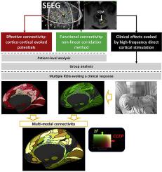
The cingulate cortex is part of the limbic system. Its function and connectivity are organized in a rostro-caudal and ventral-dorsal manner which was addressed by various other studies using rather coarse cortical parcellations. In this study, we aim at describing its function and connectivity using invasive recordings from patients explored for focal drug-resistant epilepsy. We included patients that underwent stereo-electroencephalographic recordings using intracranial electrodes in the University Emergency Hospital Bucharest between 2012-2019. We reviewed all high frequency stimulations (50 Hz) performed for functional mapping of the cingulate cortex. We used two methods to characterize brain connectivity. Effective connectivity was inferred based on the analysis of cortico-cortical potentials (CCEPs) evoked by single pulse electrical stimulation (SPES) (15 s inter-pulse interval). Functional connectivity was estimated using the non-linear regression method applied to 60 s spontaneous electrical brain signal intervals. The effective (stimulation-evoked) and functional (non-evoked) connectivity analyses highlight brain networks in a different way. While non-evoked connectivity evidences areas having related activity, often in close proximity to each other, evoked connectivity highlights spatially extended networks. To highlight in a comprehensive way the cingulate cortex's network, we have performed a bi-modal connectivity analysis that combines the resting-state broadband h2 non-linear correlation with cortico-cortical evoked potentials. We co-registered the patient's anatomy with the fsaverage FreeSurfer template to perform the automatic labeling based on HCP-MMP parcellation. At a group level, connectivity was estimated by averaging responses over stimulated/recorded or recorded sites in each pair of parcels. Finally, for multiple regions that evoked a clinical response during high frequency stimulation we combined the connectivity of individual pairs using maximum intensity projection. Connectivity was assessed by applying SPES on 2094 contact pairs and recording CCEPs on 3580 contacts out of 8582 contacts of 660 electrodes implanted in 47 patients. Clinical responses elicited by high frequency stimulations in 107 sites (pairs of contacts) located in the cingulate cortex were divided in 10 groups: affective, motor behavior, motor elementary, versive, speech, vestibular, autonomic, somatosensory, visual and changes in body perception. Anterior cingulate cortex was shown to be connected to the mesial temporal, orbitofrontal and prefrontal cortex. In the middle cingulate cortex, we located affective, motor behavior in the anterior region, and elementary motor and somatosensory in the posterior part. This region is connected to the prefrontal, premotor and primary motor network. Finally, the posterior cingulate was shown to be connected with the visual areas, mesial and lateral parietal and temporal cortex.
中文翻译:

使用颅内刺激映射的扣带皮层功能和多模式连接
扣带回皮层是边缘系统的一部分。它的功能和连接性以 rostro-caudal 和腹背方式组织,其他各种研究使用相当粗糙的皮质分割解决了这一问题。在这项研究中,我们的目标是使用对局灶性耐药性癫痫进行探索的患者的侵入性记录来描述其功能和连接性。我们纳入了 2012 年至 2019 年间在布加勒斯特大学急诊医院使用颅内电极进行立体脑电图记录的患者。我们回顾了为扣带回皮层的功能映射而执行的所有高频刺激 (50 Hz)。我们使用了两种方法来表征大脑连接。根据对单脉冲电刺激 (SPES)(15 秒脉冲间隔)诱发的皮质皮质电位 (CCEP) 的分析推断出有效连接。使用应用于 60 s 自发脑电信号间隔的非线性回归方法估计功能连接。有效的(刺激诱发的)和功能性的(非诱发的)连通性分析以不同的方式突出了大脑网络。虽然非诱发连通性证明具有相关活动的区域,通常彼此非常接近,诱发连通性突出了空间扩展的网络。为了全面突出扣带皮层网络,我们进行了双模态连接分析,将静息状态宽带 h2 非线性相关性与皮质皮质诱发电位相结合。我们将患者的解剖结构与 fsaverage FreeSurfer 模板共同注册,以执行基于 HCP-MMP 分割的自动标记。在组级别,通过对每对地块中刺激/记录或记录站点的响应进行平均来估计连通性。最后,对于在高频刺激期间引起临床反应的多个区域,我们使用最大强度投影组合了各个对的连通性。通过在 2094 个接触对上应用 SPES 并在 47 名患者中植入的 660 个电极的 8582 个接触中的 3580 个接触上记录 CCEP 来评估连接性。由位于扣带皮层中的 107 个部位(接触对)的高频刺激引起的临床反应分为 10 组:情感、运动行为、运动基本、韵律、言语、前庭、自主神经、躯体感觉、视觉和身体感知的变化。前扣带回皮层被证明与内侧颞叶、眶额叶和前额叶皮层相连。在中间扣带皮层,我们将情感、运动行为定位在前部,基本运动和躯体感觉在后部。该区域与前额叶、前运动和初级运动网络相连。最后,后扣带回与视觉区域、内侧和外侧顶叶和颞叶皮层相连。以及后部的基本运动和躯体感觉。该区域与前额叶、前运动和初级运动网络相连。最后,后扣带回与视觉区域、内侧和外侧顶叶和颞叶皮层相连。以及后部的基本运动和躯体感觉。该区域与前额叶、前运动和初级运动网络相连。最后,后扣带回与视觉区域、内侧和外侧顶叶和颞叶皮层相连。
更新日期:2020-10-01
中文翻译:
使用颅内刺激映射的扣带皮层功能和多模式连接
扣带回皮层是边缘系统的一部分。它的功能和连接性以 rostro-caudal 和腹背方式组织,其他各种研究使用相当粗糙的皮质分割解决了这一问题。在这项研究中,我们的目标是使用对局灶性耐药性癫痫进行探索的患者的侵入性记录来描述其功能和连接性。我们纳入了 2012 年至 2019 年间在布加勒斯特大学急诊医院使用颅内电极进行立体脑电图记录的患者。我们回顾了为扣带回皮层的功能映射而执行的所有高频刺激 (50 Hz)。我们使用了两种方法来表征大脑连接。根据对单脉冲电刺激 (SPES)(15 秒脉冲间隔)诱发的皮质皮质电位 (CCEP) 的分析推断出有效连接。使用应用于 60 s 自发脑电信号间隔的非线性回归方法估计功能连接。有效的(刺激诱发的)和功能性的(非诱发的)连通性分析以不同的方式突出了大脑网络。虽然非诱发连通性证明具有相关活动的区域,通常彼此非常接近,诱发连通性突出了空间扩展的网络。为了全面突出扣带皮层网络,我们进行了双模态连接分析,将静息状态宽带 h2 非线性相关性与皮质皮质诱发电位相结合。我们将患者的解剖结构与 fsaverage FreeSurfer 模板共同注册,以执行基于 HCP-MMP 分割的自动标记。在组级别,通过对每对地块中刺激/记录或记录站点的响应进行平均来估计连通性。最后,对于在高频刺激期间引起临床反应的多个区域,我们使用最大强度投影组合了各个对的连通性。通过在 2094 个接触对上应用 SPES 并在 47 名患者中植入的 660 个电极的 8582 个接触中的 3580 个接触上记录 CCEP 来评估连接性。由位于扣带皮层中的 107 个部位(接触对)的高频刺激引起的临床反应分为 10 组:情感、运动行为、运动基本、韵律、言语、前庭、自主神经、躯体感觉、视觉和身体感知的变化。前扣带回皮层被证明与内侧颞叶、眶额叶和前额叶皮层相连。在中间扣带皮层,我们将情感、运动行为定位在前部,基本运动和躯体感觉在后部。该区域与前额叶、前运动和初级运动网络相连。最后,后扣带回与视觉区域、内侧和外侧顶叶和颞叶皮层相连。以及后部的基本运动和躯体感觉。该区域与前额叶、前运动和初级运动网络相连。最后,后扣带回与视觉区域、内侧和外侧顶叶和颞叶皮层相连。以及后部的基本运动和躯体感觉。该区域与前额叶、前运动和初级运动网络相连。最后,后扣带回与视觉区域、内侧和外侧顶叶和颞叶皮层相连。


 京公网安备 11010802027423号