当前位置:
X-MOL 学术
›
Biomater. Sci.
›
论文详情
Our official English website, www.x-mol.net, welcomes your
feedback! (Note: you will need to create a separate account there.)
Ultrasound monitoring of magnet-guided delivery of mesenchymal stem cells labeled with magnetic lipid-polymer hybrid nanobubbles.
Biomaterials Science ( IF 5.8 ) Pub Date : 2020-06-12 , DOI: 10.1039/d0bm00473a Bo Zhang 1 , Xinhai Mo , Fei Yu , Yuqin Ma , Fei Yan
Biomaterials Science ( IF 5.8 ) Pub Date : 2020-06-12 , DOI: 10.1039/d0bm00473a Bo Zhang 1 , Xinhai Mo , Fei Yu , Yuqin Ma , Fei Yan
Affiliation
|
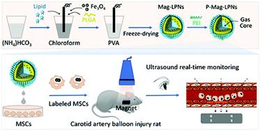
Restenosis remains a pressing clinical problem that occurs in patients undergoing revascularization procedures, such as coronary artery bypass surgery and percutaneous transluminal angioplasty. Previous reports have proved that mesenchymal stem cells (MSCs) could effectively reduce the restenosis resulting from intimal hyperplasia following vascular injury. However, non-invasive delivery of MSCs and real-time monitoring of their retention at the site of vascular injury still remain a significant challenge. Therefore, we synthesized magnetic lipid–polymer hybrid nanobubbles (Mag-LPNs) as ultrasound contrast agents for cellular labeling of MSCs, endowing these MSCs with magnetic responsibility and real-time tracking capability by ultrasound. In order to enhance the internalization efficiency of MSCs, Mag-LPNs were modified with cationic polymers to generate positively charged Mag-LPNs (P-Mag-LPNs). Intriguingly, the internalization of P-Mag-LPNs did not exhibit obvious harmful effects on the labeled MSCs in terms of cell viability and differentiation capacity. Moreover, the magnet-guided delivery of labeled MSCs in a rat carotid artery injury model developed using a 2-French balloon catheter could be tracked by ultrasound in a real-time manner. About 5-fold more MSCs were attached at the site of the injured artery with the aid of an external magnet field, compared with the absence of a magnet field. Herein, our study provides an innovative tracking platform for magnet-guided delivery of stem cells treating cardiovascular diseases.
中文翻译:

超声监测以磁性脂质-聚合物杂合纳米气泡标记的间充质干细胞的磁性引导。
再狭窄仍然是紧迫的临床问题,发生在进行血管重建手术的患者中,例如冠状动脉搭桥手术和经皮腔内血管成形术。先前的报道证明,间充质干细胞(MSCs)可有效减少血管损伤后内膜增生引起的再狭窄。然而,无创递送MSC和实时监测其在血管损伤部位的保留仍然是一个重大挑战。因此,我们合成了磁性脂质-聚合物杂化纳米气泡(Mag-LPN)作为超声造影剂,用于MSC的细胞标记,使这些MSC具有磁性和超声实时跟踪功能。为了提高MSC的内部化效率,用阳离子聚合物修饰Mag-LPN,以产生带正电的Mag-LPN(P-Mag-LPN)。有趣的是,就细胞活力和分化能力而言,P-Mag-LPN的内在化对标记的MSC没有明显的有害作用。此外,在使用2-French球囊导管开发的大鼠颈动脉损伤模型中,标记的MSC的磁引导递送可以通过超声波实时跟踪。与不存在磁场的情况相比,借助于外部磁场,大约有5倍以上的MSC附着在受伤的动脉部位。在本文中,我们的研究为磁引导干细胞治疗心血管疾病提供了创新的跟踪平台。P-Mag-LPN的内在化在细胞活力和分化能力方面对标记的MSC没有明显的有害作用。此外,在使用2-French球囊导管开发的大鼠颈动脉损伤模型中,标记的MSC的磁引导递送可以通过超声波实时跟踪。与不存在磁场的情况相比,借助于外部磁场,大约有5倍以上的MSC附着在受伤的动脉部位。在本文中,我们的研究为磁引导干细胞治疗心血管疾病提供了创新的跟踪平台。P-Mag-LPN的内在化在细胞活力和分化能力方面对标记的MSC没有明显的有害作用。此外,在使用2-French球囊导管开发的大鼠颈动脉损伤模型中,标记的MSC的磁引导递送可以通过超声波实时跟踪。与不存在磁场的情况相比,借助于外部磁场,大约有5倍以上的MSC附着在受伤的动脉部位。在本文中,我们的研究为磁引导干细胞治疗心血管疾病提供了创新的跟踪平台。在使用2-French球囊导管开发的大鼠颈动脉损伤模型中,标记的MSC的磁引导递送可以通过超声实时跟踪。与不存在磁场的情况相比,借助于外部磁场,大约有5倍以上的MSC附着在受伤的动脉部位。在本文中,我们的研究为磁引导干细胞治疗心血管疾病提供了创新的跟踪平台。使用2-French球囊导管开发的大鼠颈动脉损伤模型中标记的MSC的磁引导递送可以通过超声实时跟踪。与不存在磁场的情况相比,借助于外部磁场,大约有5倍以上的MSC附着在受伤的动脉部位。在本文中,我们的研究为磁引导干细胞治疗心血管疾病提供了创新的跟踪平台。
更新日期:2020-06-30
中文翻译:
超声监测以磁性脂质-聚合物杂合纳米气泡标记的间充质干细胞的磁性引导。
再狭窄仍然是紧迫的临床问题,发生在进行血管重建手术的患者中,例如冠状动脉搭桥手术和经皮腔内血管成形术。先前的报道证明,间充质干细胞(MSCs)可有效减少血管损伤后内膜增生引起的再狭窄。然而,无创递送MSC和实时监测其在血管损伤部位的保留仍然是一个重大挑战。因此,我们合成了磁性脂质-聚合物杂化纳米气泡(Mag-LPN)作为超声造影剂,用于MSC的细胞标记,使这些MSC具有磁性和超声实时跟踪功能。为了提高MSC的内部化效率,用阳离子聚合物修饰Mag-LPN,以产生带正电的Mag-LPN(P-Mag-LPN)。有趣的是,就细胞活力和分化能力而言,P-Mag-LPN的内在化对标记的MSC没有明显的有害作用。此外,在使用2-French球囊导管开发的大鼠颈动脉损伤模型中,标记的MSC的磁引导递送可以通过超声波实时跟踪。与不存在磁场的情况相比,借助于外部磁场,大约有5倍以上的MSC附着在受伤的动脉部位。在本文中,我们的研究为磁引导干细胞治疗心血管疾病提供了创新的跟踪平台。P-Mag-LPN的内在化在细胞活力和分化能力方面对标记的MSC没有明显的有害作用。此外,在使用2-French球囊导管开发的大鼠颈动脉损伤模型中,标记的MSC的磁引导递送可以通过超声波实时跟踪。与不存在磁场的情况相比,借助于外部磁场,大约有5倍以上的MSC附着在受伤的动脉部位。在本文中,我们的研究为磁引导干细胞治疗心血管疾病提供了创新的跟踪平台。P-Mag-LPN的内在化在细胞活力和分化能力方面对标记的MSC没有明显的有害作用。此外,在使用2-French球囊导管开发的大鼠颈动脉损伤模型中,标记的MSC的磁引导递送可以通过超声波实时跟踪。与不存在磁场的情况相比,借助于外部磁场,大约有5倍以上的MSC附着在受伤的动脉部位。在本文中,我们的研究为磁引导干细胞治疗心血管疾病提供了创新的跟踪平台。在使用2-French球囊导管开发的大鼠颈动脉损伤模型中,标记的MSC的磁引导递送可以通过超声实时跟踪。与不存在磁场的情况相比,借助于外部磁场,大约有5倍以上的MSC附着在受伤的动脉部位。在本文中,我们的研究为磁引导干细胞治疗心血管疾病提供了创新的跟踪平台。使用2-French球囊导管开发的大鼠颈动脉损伤模型中标记的MSC的磁引导递送可以通过超声实时跟踪。与不存在磁场的情况相比,借助于外部磁场,大约有5倍以上的MSC附着在受伤的动脉部位。在本文中,我们的研究为磁引导干细胞治疗心血管疾病提供了创新的跟踪平台。


 京公网安备 11010802027423号