当前位置:
X-MOL 学术
›
J. Biophotonics
›
论文详情
Our official English website, www.x-mol.net, welcomes your
feedback! (Note: you will need to create a separate account there.)
Diffuse optical assessment of cerebral-autoregulation in older adults stratified by cerebrovascular risk.
Journal of Biophotonics ( IF 2.0 ) Pub Date : 2020-06-13 , DOI: 10.1002/jbio.202000073 Ahmed A Bahrani 1, 2, 3 , Weikai Kong 1 , Yu Shang 4 , Chong Huang 1 , Charles D Smith 2, 5, 6 , David K Powell 5, 7 , Yang Jiang 2, 5, 8 , Abner O Rayapati 9 , Gregory A Jicha 2, 5, 6 , Guoqiang Yu 1
Journal of Biophotonics ( IF 2.0 ) Pub Date : 2020-06-13 , DOI: 10.1002/jbio.202000073 Ahmed A Bahrani 1, 2, 3 , Weikai Kong 1 , Yu Shang 4 , Chong Huang 1 , Charles D Smith 2, 5, 6 , David K Powell 5, 7 , Yang Jiang 2, 5, 8 , Abner O Rayapati 9 , Gregory A Jicha 2, 5, 6 , Guoqiang Yu 1
Affiliation
|
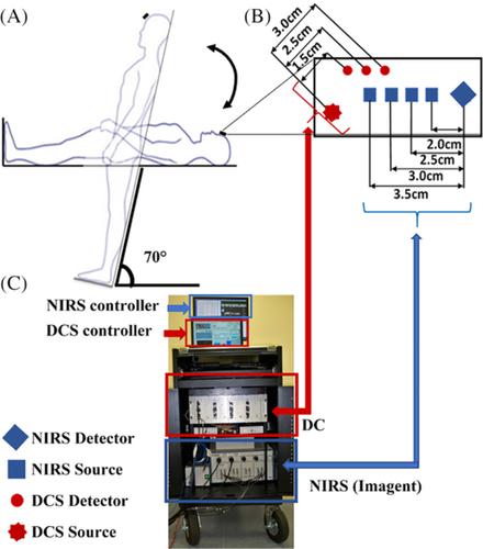
Diagnosis of cerebrovascular disease (CVD) at early stages is essential for preventing sequential complications. CVD is often associated with abnormal cerebral microvasculature, which may impact cerebral‐autoregulation (CA). A novel hybrid near‐infrared diffuse optical instrument and a finger plethysmograph were used to simultaneously detect low‐frequency oscillations (LFOs) of cerebral blood flow (CBF), oxy‐hemoglobin concentration ([HbO2]), deoxy‐hemoglobin concentration ([Hb]) and mean arterial pressure (MAP) in older adults before, during and after 70° head‐up‐tilting (HUT). The participants with valid data were divided based on Framingham risk score (FRS, 1‐30 points) into low‐risk (FRS ≤15, n = 13) and high‐risk (FRS >15, n = 11) groups for developing CVD. The LFO gains were determined by transfer function analyses with MAP as the input, and CBF, [HbO2] and [Hb] as the outputs (CA ∝ 1/Gain). At resting‐baseline, LFO gains in the high‐risk group were relatively lower compared to the low‐risk group. The lower baseline gains in the high‐risk group may attribute to compensatory mechanisms to maintain stronger steady‐state CAs. However, HUT resulted in smaller gain reductions in the high‐risk group compared to the low‐risk group, suggesting weaker dynamic CAs. LFO gains are potentially valuable biomarkers for early detection of CVD based on associations with CAs.
中文翻译:

按脑血管风险分层的老年人脑自动调节的漫反射光学评估。
早期诊断脑血管疾病 (CVD) 对于预防继发性并发症至关重要。CVD通常与异常的脑微血管系统相关,这可能会影响脑自动调节(CA)。一种新型混合近红外漫反射光学仪器和手指体积描记器用于同时检测脑血流(CBF)的低频振荡(LFO)、氧合血红蛋白浓度([HbO 2])、老年人在 70° 仰头倾斜 (HUT) 之前、期间和之后的脱氧血红蛋白浓度 ([Hb]) 和平均动脉压 (MAP)。根据 Framingham 风险评分(FRS,1-30 分)将具有有效数据的参与者分为发展为 CVD 的低风险(FRS ≤15,n = 13)和高风险(FRS >15,n = 11)组. LFO 增益由传递函数分析确定,MAP 作为输入,CBF、[HbO 2 ] 和 [Hb] 作为输出 ( CA ∝ 1/Gain). 在静息基线时,与低风险组相比,高风险组的 LFO 增益相对较低。高风险组较低的基线增益可能归因于维持更强稳态 CA 的补偿机制。然而,与低风险组相比,HUT 导致高风险组的增益减少较小,表明动态 CA 较弱。基于与 CA 的关联,LFO 增益是早期检测 CVD 的潜在有价值的生物标志物。
更新日期:2020-06-13
中文翻译:
按脑血管风险分层的老年人脑自动调节的漫反射光学评估。
早期诊断脑血管疾病 (CVD) 对于预防继发性并发症至关重要。CVD通常与异常的脑微血管系统相关,这可能会影响脑自动调节(CA)。一种新型混合近红外漫反射光学仪器和手指体积描记器用于同时检测脑血流(CBF)的低频振荡(LFO)、氧合血红蛋白浓度([HbO 2])、老年人在 70° 仰头倾斜 (HUT) 之前、期间和之后的脱氧血红蛋白浓度 ([Hb]) 和平均动脉压 (MAP)。根据 Framingham 风险评分(FRS,1-30 分)将具有有效数据的参与者分为发展为 CVD 的低风险(FRS ≤15,n = 13)和高风险(FRS >15,n = 11)组. LFO 增益由传递函数分析确定,MAP 作为输入,CBF、[HbO 2 ] 和 [Hb] 作为输出 ( CA ∝ 1/Gain). 在静息基线时,与低风险组相比,高风险组的 LFO 增益相对较低。高风险组较低的基线增益可能归因于维持更强稳态 CA 的补偿机制。然而,与低风险组相比,HUT 导致高风险组的增益减少较小,表明动态 CA 较弱。基于与 CA 的关联,LFO 增益是早期检测 CVD 的潜在有价值的生物标志物。


 京公网安备 11010802027423号