当前位置:
X-MOL 学术
›
Ann. N. Y. Acad. Sci.
›
论文详情
Our official English website, www.x-mol.net, welcomes your
feedback! (Note: you will need to create a separate account there.)
Detection and analysis of enamel cracks by ICG‐NIR fluorescence dental imaging
Annals of the New York Academy of Sciences ( IF 4.1 ) Pub Date : 2020-06-09 , DOI: 10.1111/nyas.14374 Zhongqiang Li 1 , Yoshita V Holamoge 1 , Zheng Li 1 , Waleed Zaid 2 , Michelle L Osborn 3 , Alexandra Ramos 3 , Jacob T Miller 1 , Yanping Li 4 , Shaomian Yao 3 , Jian Xu 1
Annals of the New York Academy of Sciences ( IF 4.1 ) Pub Date : 2020-06-09 , DOI: 10.1111/nyas.14374 Zhongqiang Li 1 , Yoshita V Holamoge 1 , Zheng Li 1 , Waleed Zaid 2 , Michelle L Osborn 3 , Alexandra Ramos 3 , Jacob T Miller 1 , Yanping Li 4 , Shaomian Yao 3 , Jian Xu 1
Affiliation
|
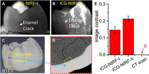
Cracked teeth are the third most common cause of tooth loss, but there is no reliable imaging tool for the diagnosis of cracks. Here, we demonstrate the feasibility of indocyanine green near‐infrared fluorescence (ICG‐NIRF) dental imaging for the detection of enamel cracks and enamel–dentin cracks in vitro in the first (ICG‐NIRF‐I, 700–950 nm) and second (ICG‐NIRF‐II, 950–1700 nm) imaging windows with transmission excitation light, and compared ICG‐NIRF with conventional NIR illumination‐II (NIRi‐II) and X‐ray imaging. Dentin cracks were detected by CT scan, while most enamel cracks, undetectable under X‐ray imaging, were clearly visible in NIR images. We found that ICG‐NIRF‐II detected cracks more effectively than NIRi‐II, and that light orientation is an important factor for crack detection: an angled exposure obtained better image contrast of cracks than parallel exposure, as it created a shadow under the crack. Crack depth could be evaluated from the crack shadow in ICG‐NIRF and NIRi‐II images; from this shadow we could determine crack depth and discriminate enamel–dentin cracks from craze lines. Cracks could be observed clearly from ICG‐NIRF images with 1‐min ICG tooth immersion, although longer ICG immersion produced images with greater contrast. Overall, our data show that ICG‐NIRF dental imaging is a useful tool for diagnosing cracked teeth at an early stage.
中文翻译:

ICG-NIR荧光牙科成像检测和分析牙釉质裂纹
牙齿开裂是牙齿缺失的第三大常见原因,但目前还没有可靠的影像工具来诊断开裂。在这里,我们证明了吲哚菁绿近红外荧光(ICG-NIRF)牙科成像在第一次(ICG-NIRF-I,700-950 nm)和第二次体外检测牙釉质裂纹和牙釉质裂纹的可行性。 (ICG-NIRF-II, 950-1700 nm) 透射激发光成像窗口,并将 ICG-NIRF 与传统 NIR 照明-II (NIRi-II) 和 X 射线成像进行比较。CT 扫描检测到牙本质裂纹,而在 X 射线成像下无法检测到的大多数牙釉质裂纹在 NIR 图像中清晰可见。我们发现ICG-NIRF-II比NIRi-II更有效地检测裂纹,并且光取向是裂纹检测的重要因素:倾斜曝光比平行曝光获得了更好的裂纹图像对比度,因为它在裂纹下产生了阴影。裂纹深度可以通过 ICG-NIRF 和 NIRi-II 图像中的裂纹阴影来评估;从这个阴影中,我们可以确定裂纹深度并将釉质-牙本质裂纹与裂纹线区分开来。从 ICG-NIRF 图像中可以清楚地观察到裂纹,ICG 牙齿浸入 1 分钟,尽管更长的 ICG 浸入产生的图像具有更大的对比度。总的来说,我们的数据表明,ICG-NIRF 牙科成像是早期诊断裂纹牙齿的有用工具。从 ICG-NIRF 图像中可以清楚地观察到裂纹,ICG 牙齿浸入 1 分钟,尽管更长的 ICG 浸入产生的图像具有更大的对比度。总的来说,我们的数据表明,ICG-NIRF 牙科成像是早期诊断裂纹牙齿的有用工具。从 ICG-NIRF 图像中可以清楚地观察到裂纹,ICG 牙齿浸入 1 分钟,尽管更长的 ICG 浸入产生的图像具有更大的对比度。总体而言,我们的数据表明,ICG-NIRF 牙科成像是早期诊断破裂牙齿的有用工具。
更新日期:2020-06-09
中文翻译:
ICG-NIR荧光牙科成像检测和分析牙釉质裂纹
牙齿开裂是牙齿缺失的第三大常见原因,但目前还没有可靠的影像工具来诊断开裂。在这里,我们证明了吲哚菁绿近红外荧光(ICG-NIRF)牙科成像在第一次(ICG-NIRF-I,700-950 nm)和第二次体外检测牙釉质裂纹和牙釉质裂纹的可行性。 (ICG-NIRF-II, 950-1700 nm) 透射激发光成像窗口,并将 ICG-NIRF 与传统 NIR 照明-II (NIRi-II) 和 X 射线成像进行比较。CT 扫描检测到牙本质裂纹,而在 X 射线成像下无法检测到的大多数牙釉质裂纹在 NIR 图像中清晰可见。我们发现ICG-NIRF-II比NIRi-II更有效地检测裂纹,并且光取向是裂纹检测的重要因素:倾斜曝光比平行曝光获得了更好的裂纹图像对比度,因为它在裂纹下产生了阴影。裂纹深度可以通过 ICG-NIRF 和 NIRi-II 图像中的裂纹阴影来评估;从这个阴影中,我们可以确定裂纹深度并将釉质-牙本质裂纹与裂纹线区分开来。从 ICG-NIRF 图像中可以清楚地观察到裂纹,ICG 牙齿浸入 1 分钟,尽管更长的 ICG 浸入产生的图像具有更大的对比度。总的来说,我们的数据表明,ICG-NIRF 牙科成像是早期诊断裂纹牙齿的有用工具。从 ICG-NIRF 图像中可以清楚地观察到裂纹,ICG 牙齿浸入 1 分钟,尽管更长的 ICG 浸入产生的图像具有更大的对比度。总的来说,我们的数据表明,ICG-NIRF 牙科成像是早期诊断裂纹牙齿的有用工具。从 ICG-NIRF 图像中可以清楚地观察到裂纹,ICG 牙齿浸入 1 分钟,尽管更长的 ICG 浸入产生的图像具有更大的对比度。总体而言,我们的数据表明,ICG-NIRF 牙科成像是早期诊断破裂牙齿的有用工具。


 京公网安备 11010802027423号