当前位置:
X-MOL 学术
›
Cytom. Part A
›
论文详情
Our official English website, www.x-mol.net, welcomes your
feedback! (Note: you will need to create a separate account there.)
Label-Free Four-Dimensional Visualization of Anaerobically Growing Electroactive Biofilms.
Cytometry Part A ( IF 2.5 ) Pub Date : 2020-06-08 , DOI: 10.1002/cyto.a.24169 Christin Koch 1 , Anne Kuchenbuch 1 , Maria Marosvölgyi 2 , Klaus Weisshart 3 , Falk Harnisch 1
Cytometry Part A ( IF 2.5 ) Pub Date : 2020-06-08 , DOI: 10.1002/cyto.a.24169 Christin Koch 1 , Anne Kuchenbuch 1 , Maria Marosvölgyi 2 , Klaus Weisshart 3 , Falk Harnisch 1
Affiliation
|
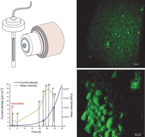
Light sheet fluorescence microscopy (LSFM) allows nondestructive, label‐free and in vivo imaging of large specimen, even at nontransparent surfaces. We show that LSFM can be applied for label‐free analyses of prokaryotes on the example of electroactive biofilms. Biofilm growth is linked to the production of current serving as measure of metabolic activity in vivo by monitoring with high spatial and temporal resolution. After 35 h of exponential growth, a homogeneous biofilm with a thickness of 9 μm was formed. This was followed by a stratification of the biofilm including the formation of 3D structures over the next 100 h. Light reflection was sufficient to visualize the biofilm structure and development over time and the terminal morphology was confirmed using fluorescence staining. This proof of concept on using LSFM for investigation of biofilms opens the door for its application in the entire field of microbial ecology. © 2020 The Authors. Cytometry Part A published by Wiley Periodicals LLC. on behalf of International Society for Advancement of Cytometry.
中文翻译:

厌氧生长的电活性生物膜的无标记四维可视化。
光片荧光显微镜 (LSFM) 允许对大型标本进行无损、无标记和体内成像,即使在不透明的表面也是如此。我们表明 LSFM 可用于以电活性生物膜为例对原核生物进行无标记分析。生物膜生长与电流的产生有关,通过高空间和时间分辨率进行监测,作为体内代谢活动的量度。指数增长 35 小时后,形成了厚度为 9 μm 的均匀生物膜。随后是生物膜的分层,包括在接下来的 100 小时内形成 3D 结构。光反射足以可视化生物膜结构和随时间的发展,并使用荧光染色确认终端形态。这种使用 LSFM 研究生物膜的概念证明为其在整个微生物生态学领域的应用打开了大门。© 2020 作者。由 Wiley Periodicals LLC 出版的Cytometry Part A。代表国际细胞计量学促进会。
更新日期:2020-07-22
中文翻译:
厌氧生长的电活性生物膜的无标记四维可视化。
光片荧光显微镜 (LSFM) 允许对大型标本进行无损、无标记和体内成像,即使在不透明的表面也是如此。我们表明 LSFM 可用于以电活性生物膜为例对原核生物进行无标记分析。生物膜生长与电流的产生有关,通过高空间和时间分辨率进行监测,作为体内代谢活动的量度。指数增长 35 小时后,形成了厚度为 9 μm 的均匀生物膜。随后是生物膜的分层,包括在接下来的 100 小时内形成 3D 结构。光反射足以可视化生物膜结构和随时间的发展,并使用荧光染色确认终端形态。这种使用 LSFM 研究生物膜的概念证明为其在整个微生物生态学领域的应用打开了大门。© 2020 作者。由 Wiley Periodicals LLC 出版的Cytometry Part A。代表国际细胞计量学促进会。


 京公网安备 11010802027423号