当前位置:
X-MOL 学术
›
Neurobiol. Aging
›
论文详情
Our official English website, www.x-mol.net, welcomes your
feedback! (Note: you will need to create a separate account there.)
Disruption of oligodendrocyte progenitor cells is an early sign of pathology in the triple transgenic mouse model of Alzheimer’s disease
Neurobiology of Aging ( IF 3.7 ) Pub Date : 2020-10-01 , DOI: 10.1016/j.neurobiolaging.2020.05.016 Ilaria Vanzulli 1 , Maria Papanikolaou 1 , Irene Chacon De-La-Rocha 1 , Francesca Pieropan 1 , Andrea D Rivera 1 , Diego Gomez-Nicola 2 , Alexei Verkhratsky 3 , José Julio Rodríguez 4 , Arthur M Butt 1
Neurobiology of Aging ( IF 3.7 ) Pub Date : 2020-10-01 , DOI: 10.1016/j.neurobiolaging.2020.05.016 Ilaria Vanzulli 1 , Maria Papanikolaou 1 , Irene Chacon De-La-Rocha 1 , Francesca Pieropan 1 , Andrea D Rivera 1 , Diego Gomez-Nicola 2 , Alexei Verkhratsky 3 , José Julio Rodríguez 4 , Arthur M Butt 1
Affiliation
|
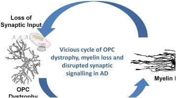
There is increasing evidence that myelin disruption is related to cognitive decline in Alzheimer's disease (AD). In the CNS, myelin is produced by oligodendrocytes, which are generated throughout life by adult oligodendrocyte progenitor cells (OPCs), also known as NG2-glia. To address whether alterations in myelination are related to age-dependent changes in OPCs, we analyzed NG2 and myelin basic protein (MBP) immunolabelling in the hippocampus of 3×Tg-AD mice at 6 and 24 months of age, compared with non-Tg age-matched controls. There was an age-related decrease in MBP immunostaining and OPC density, together with a decline in the number of OPC sister cells, a measure of OPC replication. Notably, the loss of myelin and OPC sister cells occurred earlier at 6 months in 3xTg-AD, suggesting accelerated aging, although there was not a concomitant decline in OPC numbers at this age, suggesting the observed changes in myelin were not a consequence of replicative exhaustion, but possibly of OPC disruption or senescence. In line with this, a key finding is that compared to age-match controls, OPC displayed marked morphological atrophy at 6 months in 3xTg-AD followed by morphological hypertrophy at 24 months, as deduced from significant changes in total cell surface area, total cell volume, somata volume and branching of main processes. Moreover, we show that hypertrophic OPCs surround and infiltrate amyloid-β (Aβ) plaques, a key pathological hallmark of AD. The results indicate that OPCs undergo complex age-related remodeling in the hippocampus of the 3xTg-AD mouse model. We conclude that OPC disruption is an early pathological sign in AD and is a potential factor in accelerated myelin loss and cognitive decline.
中文翻译:

少突胶质祖细胞的破坏是阿尔茨海默病三重转基因小鼠模型病理的早期征兆
越来越多的证据表明,髓鞘破坏与阿尔茨海默病 (AD) 的认知能力下降有关。在中枢神经系统中,髓鞘由少突胶质细胞产生,而少突胶质细胞在整个生命过程中由成年少突胶质祖细胞 (OPC) 产生,也称为 NG2-glia。为了解决髓鞘形成的改变是否与 OPCs 的年龄依赖性变化有关,我们分析了 6 个月和 24 个月大的 3×Tg-AD 小鼠海马中的 NG2 和髓鞘碱性蛋白 (MBP) 免疫标记,与非 Tg 相比年龄匹配的控件。MBP 免疫染色和 OPC 密度与年龄相关的下降,以及 OPC 姐妹细胞数量的下降,OPC 复制的衡量标准。值得注意的是,在 3xTg-AD 的 6 个月时,髓磷脂和 OPC 姐妹细胞的损失发生得更早,这表明衰老加速,尽管在这个年龄段 OPC 数量并没有随之下降,但这表明观察到的髓鞘变化不是复制衰竭的结果,而可能是 OPC 破坏或衰老的结果。与此一致,一个关键发现是,与年龄匹配的对照组相比,OPC 在 3xTg-AD 的 6 个月时表现出明显的形态萎缩,随后在 24 个月时出现形态肥大,这是从总细胞表面积、总细胞的显着变化推断出来的。体积,胞体体积和主要过程的分支。此外,我们发现肥大的 OPCs 包围并浸润淀粉样蛋白-β (Aβ) 斑块,这是 AD 的一个关键病理标志。结果表明 OPC 在 3xTg-AD 小鼠模型的海马中经历了复杂的与年龄相关的重塑。
更新日期:2020-10-01
中文翻译:
少突胶质祖细胞的破坏是阿尔茨海默病三重转基因小鼠模型病理的早期征兆
越来越多的证据表明,髓鞘破坏与阿尔茨海默病 (AD) 的认知能力下降有关。在中枢神经系统中,髓鞘由少突胶质细胞产生,而少突胶质细胞在整个生命过程中由成年少突胶质祖细胞 (OPC) 产生,也称为 NG2-glia。为了解决髓鞘形成的改变是否与 OPCs 的年龄依赖性变化有关,我们分析了 6 个月和 24 个月大的 3×Tg-AD 小鼠海马中的 NG2 和髓鞘碱性蛋白 (MBP) 免疫标记,与非 Tg 相比年龄匹配的控件。MBP 免疫染色和 OPC 密度与年龄相关的下降,以及 OPC 姐妹细胞数量的下降,OPC 复制的衡量标准。值得注意的是,在 3xTg-AD 的 6 个月时,髓磷脂和 OPC 姐妹细胞的损失发生得更早,这表明衰老加速,尽管在这个年龄段 OPC 数量并没有随之下降,但这表明观察到的髓鞘变化不是复制衰竭的结果,而可能是 OPC 破坏或衰老的结果。与此一致,一个关键发现是,与年龄匹配的对照组相比,OPC 在 3xTg-AD 的 6 个月时表现出明显的形态萎缩,随后在 24 个月时出现形态肥大,这是从总细胞表面积、总细胞的显着变化推断出来的。体积,胞体体积和主要过程的分支。此外,我们发现肥大的 OPCs 包围并浸润淀粉样蛋白-β (Aβ) 斑块,这是 AD 的一个关键病理标志。结果表明 OPC 在 3xTg-AD 小鼠模型的海马中经历了复杂的与年龄相关的重塑。


 京公网安备 11010802027423号